Ingredient science / Skin care
NanoStemCell
NanoStemCell™ Botanical Liposome Complex: Skin Renewal and Anti-Aging Care
Buddleja officinalis flower extract + Salvia miltiorrhiza root extract
Huzhou Puripharm Pharmaceutical Technology Co., Ltd.
Product overview · Full set of figures · November 2026
Product overview
A two-botanical ingredient blend
A water–polyol dispersion of botanical extracts and phospholipids for skin renewal, moisturization, and anti-aging formulations.
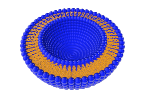
Schematic of the carrier structure. This image does not report particle-size measurements.
| Recommended use level | Formulation pH | Processing temperature |
|---|---|---|
| 0.1–5% | 4–8 | <80°C |
Ingredient origins
Botanical sources

Buddleja officinalis
Flower extract

Salvia miltiorrhiza (danshen)
Root extract
Skin biology / Formulation context
Epidermal renewal and barrier formation
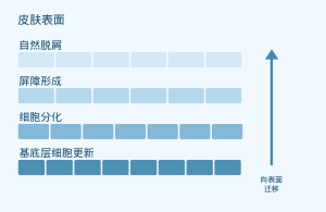
Schematic of normal epidermal physiology. It does not show results from an efficacy study of NanoStemCell.
[4] Blanpain C, Fuchs E. Nat Rev Mol Cell Biol. 2009;10:207–217. doi:10.1038/nrm2636.
Product composition
Ingredients and typical proportions
| INCI name | Range (w/w, %) | Typical proportion (w/w, %) |
|---|---|---|
| WATER | 15–35 | 32.5 |
| BUTYLENE GLYCOL | 15–35 | 30.0 |
| PROPYLENE GLYCOL | 15–35 | 32.0 |
| PHOSPHOLIPIDS | 1.5–10 | 3.5 |
| BUDDLEJA OFFICINALIS EXTRACT | 0.5–1.5 | 1.0 |
| SALVIA MILTIORRHIZA EXTRACT | 0.5–1.5 | 1.0 |
The typical composition totals 100.0%. These are supplier formulation figures and do not represent measurements of a specific batch.
Source: Supplier ingredient breakdown and composition table totaling 100%.
Carrier and production process
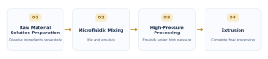
Preparing the botanical dispersion
Phospholipids act as a carrier component in the water–polyol dispersion of botanical extracts.
Research overview
Research on the botanical ingredients
| Test material | Experimental model | Research focus |
|---|---|---|
| Buddleja officinalis extract [1] | UVB-exposed HaCaT keratinocytes | NRF2-related oxidative stress responses |
| Salvianolic acid B [2] | UVB-exposed human dermal fibroblasts | MAPK/AP-1 and extracellular matrix markers |
| Salvianolic acid B [3] | Dermal fibroblasts and a mouse model of photoaging | NRF2, mitochondria, and cellular senescence |
The studies tested research extracts or isolated compounds. They do not establish the clinical efficacy of the NanoStemCell ingredient blend.
Sources: Zhu et al., 2025 [1]; Sun et al., 2015 [2]; Sun et al., 2024 [3].
Botanical ingredient research / In vitro study
Buddleja officinalis: Cellular antioxidant pathways
Experimental system
HaCaT keratinocytes; UVB: 30 mJ/cm²
Research extract, 62.5–250 µg/mL; 2-hour pretreatment
| Study measure | Direction of change |
|---|---|
| Reactive oxygen species / malondialdehyde (MDA) | Decreased |
| SOD / GSH:GSSG | Increased |
| NRF2, HO-1, and NQO1 | Stronger associated responses |
| Cell viability | Improved |
Qualitative summary. The concentrations shown apply to the extract used in this study.
[1] Zhu X, et al. Pharm Biol. 2025;63:567–581. doi:10.1080/13880209.2025.2540348.
Isolated compound research / In vitro study
Salvia miltiorrhiza: Extracellular matrix pathways
Salvianolic acid B
Purified compound, 1–100 µg/mL; UVB-exposed human dermal fibroblasts
| Pathway / measure | Study observation |
|---|---|
| MAPK / AP-1 signaling | Reduced activation |
| MMP-1 and IL-6 | Reduced expression |
| Type I procollagen and TGF-β1 | Increased |
[2] Sun Z, et al. Photochem Photobiol. 2015;91:879–886. doi:10.1111/php.12427.
Isolated compound research / Preclinical study
Salvia miltiorrhiza: NRF2 and mitochondrial function
Salvianolic acid B
UVB-exposed dermal fibroblasts and a mouse model of photoaging
NRF2 · Activation
Mitochondria · Protection
Cellular senescence · Reduction
[3] Sun JM, et al. Phytomedicine. 2024;130:155676. doi:10.1016/j.phymed.2024.155676.
Supplier images / 01 of 08
Cell colony micrographs
About these eight supplier images: Although the figure labels say NanoStemCell™, the presentation identifies Revivyl™ as the test material in the source Word document. Interpret the findings for the original test material; they are not measurements of NanoStemCell efficacy.
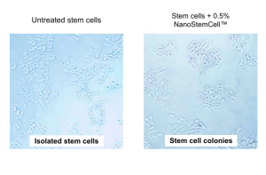
Left: untreated cells. Right: the 0.5% formulation group as labeled. The panels show isolated cells and cell colonies, respectively.
The figure labels the cells as stem cells.
Supplier images / 02 of 08
Fluorescence images of the UV response
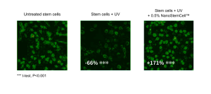
Left to right: untreated cells, UV-exposed cells, and UV-exposed cells with the 0.5% formulation shown in the label.
Original annotations: UV group −66%***; labeled formulation + UV group +171%***. *** t-test, P < 0.001.
Supplier images / 03 of 08
Ki67 histology
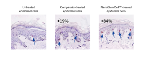
Left to right: untreated skin, comparator-treated skin, and the formulation group as labeled. Arrows indicate staining features.
Original annotations: comparator +19%; labeled formulation +84%.
Supplier images / 04 of 08
Imaging of differentiation markers
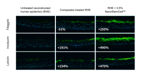
Rows: filaggrin, involucrin, and loricrin. Columns: untreated reconstructed human epidermis (RHE), comparator-treated RHE, and the 0.5% formulation group as labeled.
Original annotations (comparator / labeled formulation): filaggrin −51% / +250%; involucrin +191% / +490%; loricrin +154% / +470%.
Supplier images / 05 of 08
KLK5 immunostaining
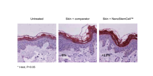
Left to right: untreated skin, comparator-treated skin, and the formulation group as labeled. The image retains the KLK5 staining and the supplier’s original numerical annotations.
Original annotations: comparator −9%; labeled formulation +13%. Figure note: * t-test, P < 0.05.
Supplier images / 06 of 08
Histology of surface desquamation
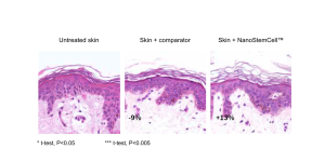
Left to right: untreated skin, comparator-treated skin, and the formulation group as labeled. Numerical annotations are retained as shown in the source.
Original annotations: comparator −9%; labeled formulation +13%. Figure notes: * t-test, P < 0.05; *** t-test, P < 0.005.
Supplier images / 07 of 08
OCT images over time
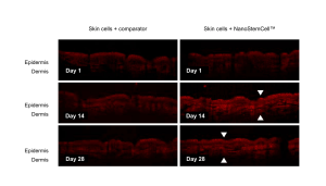
Columns: comparator and the formulation group as labeled. Rows: days 1, 14, and 28. Arrows are retained as shown in the source.
Skin layers labeled in the image: epidermis and dermis.
Supplier images / 08 of 08
Photographs of silicone replicas of crow’s feet
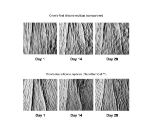
Top row: comparator. Bottom row: the formulation group as labeled. In each row, replicas are shown from left to right for days 1, 14, and 28.
Application development
Skin care formulation applications
01 · Daily serums
Formulations for skin conditioning and improved skin texture.
02 · Day and night creams
Facial moisturization and anti-aging formulations.
03 · Body lotions for dry skin
Lotion formats for daily face and body care.
04 · Water-based skin care mists
Aqueous formats for daily skin care.
Formulation guidance
Recommended incorporation conditions
0.1–5.0% · Of the finished formulation
| Item | Recommendation |
|---|---|
| Temperature | Below 80°C |
| Formulation pH | 4.0–8.0 |
| Mixing | Gentle stirring |
Add near the end of processing or to the aqueous phase.
Verify compatibility, stability, and performance in the finished formulation at the intended use level.
Use-level calculations
Ingredient contributions at different use levels
Estimated component concentrations in the finished formulation (w/w, %)
| NanoStemCell use level | 0.1% | 1.0% | 3.0% | 5.0% |
|---|---|---|---|---|
| Buddleja officinalis extract | 0.001 | 0.010 | 0.030 | 0.050 |
| Salvia miltiorrhiza extract | 0.001 | 0.010 | 0.030 | 0.050 |
| Phospholipids | 0.0035 | 0.035 | 0.105 | 0.175 |
| Total polyols¹ | 0.062 | 0.620 | 1.860 | 3.100 |
¹ Sum of propylene glycol and butylene glycol. Calculation: ingredient use level × typical proportion of the component.
Technical information
Quality specifications and storage
| Quality parameter | Specification |
|---|---|
| Appearance | Pale yellow to yellow liquid |
| Total aerobic count | ≤100 CFU/g |
| Yeasts and molds | ≤10 CFU/g |
| Escherichia coli / Salmonella | Not detected per g |
Storage temperature · 4–25°C
Shelf life in unopened original packaging · 12 months
Protect from sunlight. Use plastic containers.
Research and product materials
References and source documents
Published research
[1] Zhu X, et al. Pharm Biol. 2025;63(1):567–581.doi:10.1080/13880209.2025.2540348
[2] Sun Z, et al. Photochem Photobiol. 2015;91(4):879–886.doi:10.1111/php.12427
[3] Sun JM, et al. Phytomedicine. 2024;130:155676.doi:10.1016/j.phymed.2024.155676
[4] Blanpain C, Fuchs E. Nat Rev Mol Cell Biol. 2009;10(3):207–217.doi:10.1038/nrm2636
Product source documents
Puripharm NanoStemCell product information, revision jv2; English product materials; supplier image set (nanostemcell_pic180709); ingredient composition table; origin statement; process documents; technical data; and certificate of analysis (COA) dated December 23, 2025.
Huzhou Puripharm Pharmaceutical Technology Co., Ltd.
NanoStemCell
Botanical research to support skin care formulation development
www.puriactives.com
service@puripharm.com | +86-572-2745768
Building 6, No. 1366 Hongfeng Road, Huzhou, Zhejiang, China