ENGLISH VERSION
NanoActive Alavelle™Aminolevulinic Acid Nanoliposomes
Product overview
NanoActive Alavelle™
Aminolevulinic Acid Nanoliposomes · Advanced ALA Delivery for Skin and Scalp Innovation
Powered by PURISOME® FlexVes™ Liposome Technology
Purui Biomedical Technology Co., Ltd. · Innovation for Health and Beauty
Product identity
One active ingredient, engineered for delivery
Product name
NanoActive Alavelle™ Aminolevulinic Acid Nanoliposomes
Active ingredient INCI name
AMINOLEVULINIC ACID
Scientific identity
5-Aminolevulinic acid (5-ALA)
Delivery platform
PURISOME® FlexVes™ Liposome Technology
AMINOLEVULINIC ACID identifies the active component. The complete raw material is a multicomponent liposomal composition; see the technical documentation for the full INCI list.
5-ALA is a natural biochemical precursor of protoporphyrin IX (PpIX) in the heme pathway.
Application development landscape
■ Sprays for facial and scalp coverage
■ Topical aqueous gels
■ Scalp serums and tonics
■ Professional photoactivated treatments
■ Daylight photodynamic applications
■ Oral formulation development
Why this ingredient, why now
Development opportunities
5-ALA is one of the most extensively studied actives in photodynamic dermatology—and one of the most challenging to formulate.
4 Interconnected opportunities, one delivery platform
01 Stabilize the active. Protect a pH-sensitive hydrophilic molecule within engineered vesicles, turning aqueous stability measured in days into a usable shelf life.
02 Deliver where it matters. Improve transport to target skin compartments and sebum-rich follicles, rather than leaving the active on the surface.
One platform, multiple formats. Sprays, gels, scalp care, professional photoactivation and daylight photodynamic applications, with a separate oral development pathway.
Design for comfort. Build dose, incubation and light exposure into the protocol so efficacy and tolerability are developed together.
Mechanism of action
ALA in cell biology: the heme pathway

What happens when ALA is supplied externally?
Exogenous ALA bypasses the rate-limiting ALA synthase step and heme feedback control. In metabolically active cells, including sebaceous glands and proliferative epidermis, limited ferrochelatase capacity favors PpIX accumulation in target tissue.
Schematic of heme biosynthesis. 5-ALA is a metabolic precursor of PpIX; it is not itself a photoactivated photosensitizer.
Innovation pathways
Two routes to application innovation
Route A—photoactivated chemistry
Vesicular delivery → intracellular PpIX → controlled illumination → photodynamic response
Skin renewal and photorejuvenation: 0.5% liposomal ALA spray + IPL has been studied for wrinkles and photodamage.
Pilosebaceous care: follicular delivery combined with IPL has been studied in inflammatory acne.
Daylight protocols: continuous low-dose activation has clinical validation in actinic keratosis studies.
Piccioni et al. | J Dermatolog Treat | 2011 · Yeung et al. | Dermatol Surg | 2011 · Dirschka et al. | JEADV | 2019
Route B—non-photodynamic combinations
ALA + iron/peptide cofactors → dermal papilla signaling → follicle research
No light required. ALA combined with ferric ammonium citrate activates ERK/AKT signaling in dermal papilla cells and supports hair-shaft elongation in ex vivo human follicles.
Combination logic. Peptide (GHK) and iron partners define the studied systems; results belong to the combination, not to ALA alone.
Ryu et al. | FEBS Open Bio | 2026 · Lee et al. | Ann Dermatol | 2016 · Morokuma et al. | Int J Dermatol | 2008
Formulation science
Why formulation matters: ALA is highly formulation-sensitive
1 · A hydrophilic zwitterion facing a lipid barrier
At physiological pH, ALA carries both positive and negative groups and partitions poorly into the lipid-rich stratum corneum. Unformulated aqueous ALA largely remains on the surface.
2 · pH-triggered dimerization, followed by oxidation
Above approximately pH 5, the deprotonated amino group of one ALA molecule attacks another molecule’s ketone group. The resulting dihydropyrazine (DHPY) oxidizes in air to inactive pyrazine (PY), with loss of PpIX-forming activity.
3 · Second-order decay that antioxidants cannot stop
Degradation accelerates with the square of the free ALA concentration. Chelators and antioxidants cannot stop the primary pathway; effective controls are pH, temperature and oxygen exclusion.
Measured half-life of 1% aqueous ALA (50 °C)
3 h
pH 7.42—near physiological pH
257 h
pH 4.81—mild acidification
37 d
Stable at pH 2.35—fully protonated amino group
The active’s microenvironment—pH, oxygen and molecular mobility—determines shelf life. Formulation creates the challenge and provides the solution.
Bunke et al. | J Pharm Sci | 2000 | PMID: 10980508 · Elfsson et al. | Eur J Pharm Sci | 1999 | PMID: 9845784
Delivery platform
PURISOME® FlexVes™: an architecture for hydrophilic actives
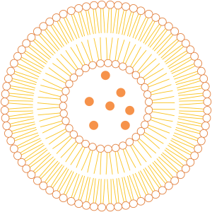
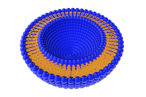
Phospholipid bilayer membrane
Aqueous core—hydrophilic ALA payload
Flexible, skin-compatible vesicle design
What this architecture does
Accommodates the payload correctly. ALA resides in aqueous and interfacial regions, associating with phospholipid headgroups rather than dissolving in oil.
Stabilizes the chemistry. Restricted molecular mobility and controlled local pH slow the dimerization that degrades aqueous ALA.
Prepares for delivery. A deformable lipid membrane supports partitioning into the stratum corneum and sebum-rich follicles.
Published studies of liposomal ALA delivery and treatment protocols are cited as technical background for this platform class.
PURISOME® FlexVes™ Liposome Technology
Formulation science
Stability through formulation design
01 Interfacial association. ALA associates with phospholipid headgroups, reducing free-molecule collisions that drive second-order dimerization.
02 Microenvironmental pH. A mildly acidic interface keeps amino groups protonated and unable to attack neighboring ketone groups.
03 Compartmentalization. Interfacial membranes restrict orientation and contact, turning one bulk phase into many isolated microenvironments.
04 Locally reduced oxygen. The lipid environment limits oxygen access to DHPY intermediates, slowing irreversible oxidation to inactive PY.
Published precedent. In a phospholipid nanovesicular gel containing 7.8% ALA, reported association with phosphatidylcholine polar headgroups extended stability from days in water to a shelf life of 24–36 months. PURISOME® vesicles apply the same physical rationale.
Days Aqueous solution → 24–36 months
Reported shelf life of a 7.8% ALA nanovesicular gel
Quality note: measure chemical stability (ALA content and pyrazine impurities) separately from vesicle size and appearance. Colloidal stability alone does not prove chemical stability. Antioxidants have little effect on the main condensation pathway; effective measures include pH below approximately 5 and oxygen exclusion.
Reinhold | Future Oncol | 2017 | PMID: 28805092 · Bunke et al. | J Pharm Sci | 2000 | PMID: 10980508
Formulation science
Aqueous vs nanovesicular systems: what changes for ALA?
Table expanded in original row order; all column labels and data are retained.

Read the shelf-life row correctly: 24–36 months refers to that specific proprietary nanovesicular gel. It is neither a general emulsion claim nor a NanoActive Alavelle™ specification. Every formulation needs its own stability program with ALA content and pyrazine impurities as endpoints; size and appearance alone cannot prove chemical stability.
Bunke et al. | J Pharm Sci | 2000 | PMID: 10980508 · Reinhold | Future Oncol | 2017 | PMID: 28805092 · Elfsson et al. | Eur J Pharm Sci | 1999
Delivery evidence
Delivery into skin and follicular compartments
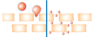
Schematic: smaller vesicles can pass between corneocytes

Confocal cross-sections: as particle size decreases (a → c), fluorescent material reaches deeper skin layers
Particle size defines the depth budget
Vesicle size and membrane deformability determine how far the carrier delivers its payload. Comparative studies show that deformable liposomes can substantially improve ALA delivery into skin.
Follicles provide a fast route
Hair follicles and sebaceous glands bypass the stratum corneum and extend into the dermis. Lipid vesicles readily partition into this sebum-rich environment.
Local retention rather than systemic absorption
The goal is retention in skin compartments where PpIX forms. After topical ALA application, plasma levels remained within normal reference ranges; delivery is intended to be local.
Oh et al. | Eur J Pharm Sci | 2011 | PMID: 21782942 · Schmitz et al. | Photodiagn Photodyn Ther | 2016 · Rick et al. | J Photochem Photobiol B | 1997
Evidence—delivery
Low-concentration liposomal ALA: rapid, self-limiting PpIX formation
A 0.5% liposomal ALA spray saturates skin fluorescence in approximately 2 hours, with clearance within 8 hours.
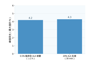
Mean peak skin PpIX fluorescence, expressed as a percentage of maximum (healthy volunteers, n = 10). The source describes the protocols as statistically equivalent (P = 0.47).
Plateau at about 2 h. Non-occluded 0.5% liposomal ALA reaches a fluorescence plateau after approximately two hours.
Return to baseline within 8 h. The signal declines within minutes after removal and clears fully within eight hours, giving a short photosensitivity window.
Comparison with 20% cream. Fluorescence continues rising for approximately eight hours after removal of the high-concentration cream, producing a longer, less controllable exposure tail.
Concentrations here describe dose and kinetics, not a fold-increase in bioavailability.
Christiansen et al. | Lasers Surg Med | 2007 | PMID: 17457834
Mechanism—light
Photoactivation: PpIX photochemistry needs photons and oxygen
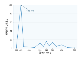
PpIX absorption profile: Soret band (approximately 405 nm) and Q bands (approximately 510–635 nm). Schematic, not to scale.
Blue light, 405–417 nm
Red light, 630–635 nm
Daylight and IPL cover the full activation spectrum
1 · Excitation. PpIX absorbs a photon and enters a long-lived triplet state.
2 · Energy transfer. Triplet PpIX transfers energy to dissolved molecular oxygen (Type II photochemistry), producing singlet oxygen and ROS.
3 · Local response. ROS act within micrometers of PpIX, mapping the response to the tissue reached by delivery.
4 · Dose rate is a design variable. Spectrum, irradiance and duration shape efficacy and comfort. Continuous low-rate daylight activation can deliver a comparable cumulative dose with much less pain than high-intensity lamps.
For photochemistry and PpIX photophysics, see Bunke et al. | J Pharm Sci | 2000 and Christiansen et al. | Lasers Surg Med | 2007.
Evidence—skin renewal
Photodynamic photorejuvenation: a measurable light dose guided by the skin
The liposomal ALA concept was developed for photodamaged skin: deliver a low-dose precursor, use skin fluorescence to indicate PpIX formation, then activate it with light.
1.7 vs 1.1
Mean participant-rated improvement in skin quality in a fluorescence-guided photodynamic rejuvenation study (0–4 scale)
2007
Mapping the fluorescence kinetics of 0.5% liposomal ALA in normal skin
2009
Fluorescence-controlled photorejuvenation: skin signals determine the light dose
2011
Wrinkle study: 30 participants, three liposomal ALA + IPL sessions
2011
A pilot study in inflammatory facial acne using the same spray + IPL approach
Implication for development: spray format, a 0.5% dose and IPL use have been published together, providing a literature template for professional photoactivated skin care.
Christiansen et al. | Lasers Surg Med | 2007 · Bjerring et al. | Lasers Surg Med | 2009; 41:327–336 · Piccioni et al. | 2011 · Yeung et al. | 2011
Clinical evidence—photorejuvenation
Wrinkle scores fall after three 0.5% liposomal ALA + IPL sessions
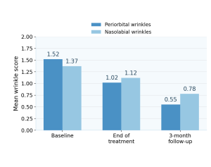
Mean modified Fitzpatrick wrinkle scores (lower is milder). These are means on an ordinal scale, not direct percentage measurements of wrinkle depth.
Study design
30 participants with facial photodamage
Three sessions of 0.5% liposomal ALA spray + IPL
Three-week intervals
Follow-up three months after the final session
Scores improved further between the last session and follow-up, consistent with ongoing post-treatment remodeling.
Piccioni et al. | J Dermatolog Treat | 2011 | DOI: 10.3109/09546634.2011.590791 | PMID: 21801099
Clinical evidence—dermal remodeling
Beyond wrinkle scores: the procollagen response after ALA-PDT
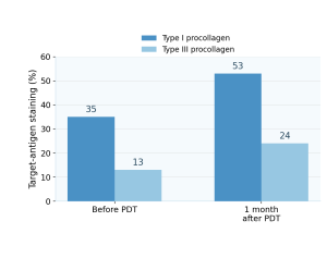
Mean target-antigen staining (%), paired biopsies before and one month after two ALA-PDT sessions. Values are approximate readings from Figure 2C. This is a photoactivation protocol, not evidence for unilluminated ALA.
Original histology (Figure 2B): Masson’s trichrome staining of dermal collagen

Study protocol
Two ALA-PDT sessions one month apart; 14 patients with facial actinic keratoses; 25 paired biopsies before treatment and one month afterward.
Histological findings
↑ Total dermal collagen; ↑ type I/III procollagen
↑ Epidermal TGF-β and TGF-β receptor II
↓ MMP-1, MMP-3 and MMP-12 (collagen-degrading enzymes)
↓ Solar elastosis; ↓ inflammatory infiltrates
ALA-PDT → ↑ TGF-β → ↑ procollagen I/III → ↓ MMP-1/3/12 → ↓ elastosis
Park MY, Sohn S, Lee ES, Kim YC | J Am Acad Dermatol | 2010;62:85–95 | DOI: 10.1016/j.jaad.2009.06.025 | PMID: 19926165
Clinical evidence—acne
Follicular delivery supports improvement in inflammatory acne
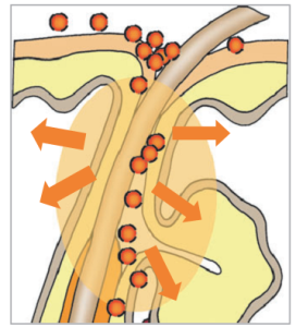
Pilosebaceous unit: the hair follicle and sebaceous gland as delivery targets
Why follicles matter. Inflammatory acne is a disease of the pilosebaceous unit, a structure preferentially accessed by lipid vesicles.
Delivery rationale. The lipophilic vesicle surface mixes with sebum, carrying the hydrophilic precursor down the follicular canal.
Activation rationale. PpIX forms in sebaceous tissue; IPL then drives a local photodynamic response.
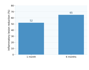
Mean reduction in inflammatory lesion counts (%) after 0.5% liposomal ALA spray + IPL; pilot study in 12 participants.
Yeung et al. | Dermatol Surg | 2011 | DOI: 10.1111/j.1524-4725.2011.01929.x | PMID: 21414073
Evidence—tissue level
Tissue evidence: PpIX forms where the carrier delivers ALA
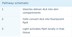
Full-thickness ex vivo skin—Maisch 2010
An ALA nanoemulsion induced measurable PpIX fluorescence in a full-thickness skin model: the delivered precursor reached viable epidermis and was metabolized there.
Ex vivo human skin comparison—Schmitz 2016
Formulation choice changed both epidermal ALA delivery and PpIX formation. The carrier, as well as the active, determines the tissue dose.
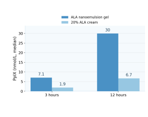
PpIX in tissue lysates (nmol/L, median), ex vivo human eyelid skin from 10 donors; redrawn from the reported data (Schmitz et al., 2016).
Maisch et al. | Exp Dermatol | 2010 | DOI: 10.1111/j.1600-0625.2009.01001.x · Schmitz et al. | Photodiagn Photodyn Ther | 2016 | DOI: 10.1016/j.pdpdt.2015.11.004
Evidence—imaging
Imaging evidence: fluorescence follows the carrier, not just the active
Schmitz 2016, Figure 2—original image
Placebo
Nanoemulsion gel
20% cream
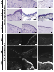
Image labels: 3 h, 6 h and 12 h; hematoxylin & eosin; PpIX fluorescence. Columns: placebo, nanoemulsion gel, 20% cream.
H&E and PpIX fluorescence cryosections; ex vivo human eyelid skin from 10 donors.
Christiansen 2007, Figure 6—original image
Follicle/lesion-selective signal (A) vs uniform signal (B)
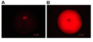
Measured skin-surface PpIX fluorescence: 0.5% liposomal ALA spray for 3 h without occlusion (A) vs occluded 20% ALA cream (B). The lipid carrier concentrates the signal in follicles and lesions.
de Leeuw 2010, Figure 3—original image
Split-face comparison, one day after identical IPL settings
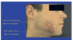
Image labels: 0.5% 5-ALA liposomes; 20% 5-ALA cream; after IPL treatment.
Non-occluded 0.5% liposomal ALA spray (left circled area) vs occluded 20% ALA cream (right). Tolerability differs with delivery and protocol, rather than with the active alone.
Original images reproduced from the cited publications for scientific reference. Third-party trademarks appearing in the source images were neutralized in the supplied PPT. Microscopy supports localization, not quantitative depth measurement.
Schmitz et al. | Photodiagn Photodyn Ther | 2016 | DOI: 10.1016/j.pdpdt.2015.11.004 · Christiansen et al. | Lasers Surg Med | 2007 | PMID: 17457834 · de Leeuw et al. | JEADV | 2010 | DOI: 10.1111/j.1468-3083.2009.03447.x
Protocol design
Designing for efficacy and comfort together
Table expanded in original row order; all column labels and data are retained.

Comfort is a property of the whole protocol—formulation, incubation and illumination—not encapsulation alone.
Christiansen et al. | Lasers Surg Med | 2007 | PMID: 17457834 · Dirschka et al. | JEADV | 2019 | PMID: 30022544
Daylight photodynamic applications
Daylight as the light source: a simple, continuous protocol
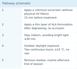
Why continuous low-intensity light can remain comfortable
During two hours of daylight, PpIX is activated and photobleached almost as soon as cells synthesize it. Tissue PpIX stays at a low steady state, delivering cumulative photochemical exposure without the concentration spikes associated with treatment pain.
Protocol flexibility: seasonal natural daylight or simulated-daylight lamps that avoid weather and latitude constraints, both evaluated clinically.
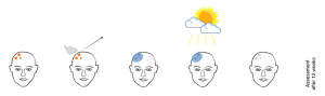
Image label: assessment after 12 weeks.
Original daylight PDT treatment schematic (Wiegell et al., 2008, Figure 1). A professional clinical protocol, not consumer sun-exposure guidance.
Schematic: Wiegell et al. | Br J Dermatol | 2008 | PMID: 18294318 · Protocol: Dirschka et al. | JEADV | 2019 | PMID: 30022544 · Simulated daylight: Bai-Habelski et al. | 2021
Clinical evidence—daylight
Daylight PDT: comparable clearance, lower recurrence and very little pain
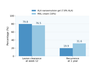
Values are percentages. Single-session daylight PDT; randomized intraindividual controlled phase III study in 52 patients with actinic keratoses. Non-inferiority P < 0.0001; recurrence difference P = 0.01.
1.2 vs >4
Mean maximum pain during daylight exposure (VAS 0–10), compared with reported mean values above 4 for conventional lamp-based PDT
Reading the evidence correctly
Indication: actinic keratosis, a clinical endpoint shown here as delivery-platform evidence, not cosmetic efficacy.
Carrier: a phospholipid nanoemulsion gel, related to liposomal technology, clinically demonstrating daylight-compatible ALA delivery.
Design: randomized split-face non-inferiority study; 52 randomized, 49 per protocol; gel = 10% ALA hydrochloride (approximately 7.8% free acid), versus 16% methyl aminolevulinate cream.
Dirschka et al. | J Eur Acad Dermatol Venereol | 2019 | DOI: 10.1111/jdv.15185 | PMID: 30022544
Scalp science
Follicle biology: a second home for vesicular delivery
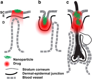
Image labels: nanoparticle, drug; SC = stratum corneum, E = epidermis, D = dermis; dermal–epidermal junction; blood vessel.
Particle-delivery pathways at the follicle: surface film, follicular canal and deeper follicular penetration
The follicle is a living, cycling mini-organ
Dermal papilla cells at its base drive the anagen–catagen–telogen cycle. Their metabolic activity makes them a relevant target for active combinations.
Follicles bypass the barrier
The follicular canal extends into the dermis, with no mature stratum corneum in deeper regions—a natural bypass available to vesicles.
Sebum supports lipid vesicles
The sebum-rich canal is chemically compatible with phospholipid vesicles, supporting retention and release near the papilla.
This development route requires no photoactivation: the non-photodynamic track for NanoActive Alavelle™.
Scalp science
Inside the papilla: ERK/AKT signaling triggered by ALA + iron
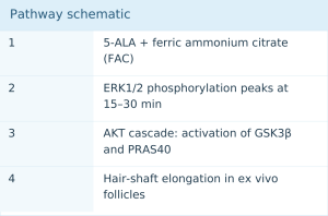
HMOX1 (HO-1) remained unchanged. Signaling involved ERK/AKT, rather than the sometimes-assumed ALA + iron heme oxygenase pathway.
Reading the evidence correctly
A combination, not ALA alone. Cellular and follicular responses were observed with joint 5-ALA and iron exposure and cannot be attributed to ALA itself.
Models, not patients. Immortalized dermal papilla cells and ex vivo follicle organ culture are mechanistic models. Clinical endpoints require clinical studies.
Ryu et al. | FEBS Open Bio | 2026 | DOI: 10.1002/2211-5463.70230 | PMID: 41873782
Scalp science
Original data from follicle models (open access)
Phospho-kinase array—Figure 3A (original)
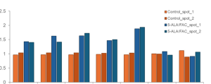
Image labels: Control_spot_1/2 and 5-ALA/FAC_spot_1/2. The supplied PPT image has no visible x-axis item names; none have been inferred.
Relative phosphorylation in dermal papilla cells, 1 µM 5-ALA/FAC vs control: increased kinases along the ERK/AKT axis.
Follicle organ culture—Figure 4B (original)
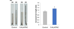
Image labels: D0/D5 = days 0/5; control; 5-ALA/FAC combination; increased hair length (mm). The asterisk is the original significance marker; P < 0.05 is specified in the text.
Hair-shaft elongation after five days of ex vivo human follicle organ culture, combination vs control, n = 20, P < 0.05.
Original images reproduced under CC BY 4.0 (© 2026 the authors; FEBS Open Bio / John Wiley & Sons). Results belong to the 5-ALA + FAC combination, not ALA alone, and to model systems rather than human clinical outcomes.
Ryu et al. | FEBS Open Bio | 2026;16:1527–1536 | DOI: 10.1002/2211-5463.70230 | PMID: 41873782 | Open access CC BY 4.0
Scalp science
Three levels of hair evidence: cells, follicles and humans
Table expanded in original row order; all column labels and data are retained.

What the three studies have in common
Every positive hair-related result combined ALA with an iron source or peptide partner: the combination is the active principle. None used photoactivation; this is a chemistry-and-delivery pathway, not PDT.
Ryu et al. | FEBS Open Bio | 2026 | PMID: 41873782 · Lee et al. | Ann Dermatol | 2016 | PMID: 27489425 · Morokuma et al. | Int J Dermatol | 2008 | PMID: 19126021
Product formats
Sprays and aqueous gels: built around vesicles
Leave-on spray
Forms a thin, even film without rubbing, treating the application site gently. Covers large areas and reaches the scalp through hair. Low viscosity naturally suits aqueous vesicle dispersions.
Precedent: the published liposomal ALA skin studies used spray formulations.
Aqueous gel
Provides longer residence at precisely targeted sites, with accurate, repeatable dosing.
A water-based gel keeps hydrophilic ALA in a compatible environment while vesicles handle delivery.
Formulation guardrails from the stability literature
pH approximately 5–6. Balance ALA chemical stability with skin compatibility.
Protect from oxygen and light. Packaging is part of the stability strategy.
Test chemistry, not just colloids. Track ALA content and pyrazine condensation impurities; size and PDI alone cannot establish active integrity.
Watch cosolvent levels. Published ALA nanoemulsion data associate excess polyols with impurity formation.
Bunke et al. | J Pharm Sci | 2000 | PMID: 10980508 · Christiansen et al. | Lasers Surg Med | 2007 | PMID: 17457834
Scalp science
Scalp-care concepts: the non-photodynamic route
Leave-on scalp serum
A vesicle dispersion targeting the follicular canal, designed for overnight residence near the papilla’s “doorway.”
Daily nourishing mist
A lightweight aqueous formula provides broad, even coverage through hair, using a format with published precedent in ALA scalp applications.
Pretreatment gel
Controlled, timed contact before rinsing, for consumers who prefer a defined exposure window.
Combination-first design
Published hair-study results belong to ALA combined with iron or peptide partners. Formulate the concept around the combination, not ALA alone.
Concepts, not specifications. These are development directions for brand partners. Concentration, packaging and shelf life depend on each partner’s formulation and stability studies. None of these concepts involves photoactivation.
Basis: Ryu et al. | FEBS Open Bio | 2026 | PMID: 41873782 · Lee et al. | Ann Dermatol | 2016 | PMID: 27489425 · Morokuma et al. | Int J Dermatol | 2008 | PMID: 19126021
Professional applications
Photoactivated formats: formulation, light and evidence
Table expanded in original row order; all column labels and data are retained.

How Purui Biomedical positions this evidence
These are professional protocols studied in clinical settings. For NanoActive Alavelle™, they provide platform evidence that low-concentration ALA in engineered carriers works under real illumination, rather than cosmetic product claims.
Piccioni et al. | J Dermatolog Treat | 2011 | PMID: 21801099 · Yeung et al. | Dermatol Surg | 2011 | PMID: 21414073 · Dirschka et al. | JEADV | 2019 | PMID: 30022544 · Bai-Habelski et al. | Photodiagn Photodyn Ther | 2021 | DOI: 10.1016/j.pdpdt.2021.102692
Route-specific science
Another route: lessons from oral ALA pharmacokinetics
≈60%
Oral 5-ALA bioavailability in healthy volunteers
≈45 min
Elimination half-life—rapid systemic clearance
Rapid absorption, onset and clearance: kinetics fundamentally different from a topical leave-on product.
Route-specific data cannot be transferred across routes
No cross-route extrapolation. Oral bioavailability and clearance do not establish topical penetration, and vesicular skin-delivery data do not establish oral exposure.
A separate development program is required. Any oral concept based on ALA chemistry needs its own regulatory and clinical pathway. The PURISOME® platform discussed here targets topical delivery.
Dalton et al. | J Pharmacol Exp Ther | 2002 | DOI: 10.1124/jpet.301.2.507 | PMID: 11961050
Formulator’s checklist
Six decisions that determine whether ALA survives and performs
Table expanded in original row order; all column labels and data are retained.

Elfsson et al. | Eur J Pharm Sci | 1999 | PMID: 9845784 · Bunke et al. | J Pharm Sci | 2000 | PMID: 10980508 · Dalton et al. | J Pharmacol Exp Ther | 2002 | PMID: 11961050
Platform overview
One platform, four evidence-informed directions
Table expanded in original row order; all column labels and data are retained.

Each evidence label corresponds to a full citation in the reference section. NanoActive Alavelle™ product specifications are provided by Purui Biomedical under project-specific agreements.
Closing perspective
One molecule, one platform, multiple thoughtfully designed formats.
01 Stabilize the chemistry
Control pH and oxygen; demonstrate integrity by testing ALA and its condensation impurities.
02 Deliver where it matters
Flexible phospholipid vesicles carry hydrophilic ALA across the barrier and along the follicular bypass.
03 Design for comfort
Low loading, short contact and gentle illumination: comfort is a protocol property.
Purui Biomedical Technology Co., Ltd.
NanoActive Alavelle™ Aminolevulinic Acid Nanoliposomes—powered by PURISOME® Liposome Technology
Innovation for Health and Beauty
Regulatory statement: finished products containing NanoActive Alavelle™ must comply with the laws and regulations of the countries or regions where they are manufactured and sold. The brand is responsible for final-product regulatory compliance.
References
References (Part 1 of 2)
01 · Rick K, Sroka R, Stepp H, Kriegmair M, Huber RM, Jacob K, Baumgartner R. Pharmacokinetics of 5-aminolevulinic acid-induced protoporphyrin IX in skin and blood. J Photochem Photobiol B. 1997;40(3):313–319. DOI: 10.1016/S1011-1344(97)00076-6 · PMID: 9372622
02 · Elfsson B, Wallin I, Eksborg S, Rudaeus K, Ros AM, Ehrsson H. Stability of 5-aminolevulinic acid in aqueous solution. Eur J Pharm Sci. 1999;7(2):87–91. DOI: 10.1016/S0928-0987(98)00098-8 · PMID: 9845784
03 · Bunke A, Zerbe O, Schmid H, Burmeister G, Merkle HP, Gander B. Degradation mechanism and stability of 5-aminolevulinic acid. J Pharm Sci. 2000;89(10):1335–1341. PMID: 10980508
04 · Dalton JT, Yates CR, Yin D, Straughn A, Marcus SL, Golub AL, Meyer MC. Clinical pharmacokinetics of 5-aminolevulinic acid in healthy volunteers and patients at high risk for recurrent bladder cancer. J Pharmacol Exp Ther. 2002;301(2):507–512. DOI: 10.1124/jpet.301.2.507 · PMID: 11961050
05 · Christiansen K, Bjerring P, Troilius A. 5-ALA for photodynamic photorejuvenation — optimization of treatment regime based on normal-skin fluorescence measurements. Lasers Surg Med. 2007;39(4):302–310. PMID: 17457834
06 · Morokuma Y, Yamazaki M, Maeda T, Yoshino I, Ishizuka M, Tanaka T, Ito Y, Tsuboi R. Hair growth stimulatory effect by a combination of 5-aminolevulinic acid and iron ion. Int J Dermatol. 2008;47(12):1298–1303. PMID: 19126021
07 · Bjerring P, Christiansen K, Troilius A, Bekhor P, de Leeuw J. Skin fluorescence controlled photodynamic photorejuvenation (wrinkle reduction). Lasers Surg Med. 2009;41(5):327–336.
08 · Maisch T, Santarelli F, Schreml S, Babilas P, Szeimies RM. Fluorescence induction of protoporphyrin IX by a new 5-aminolevulinic acid nanoemulsion used for photodynamic therapy in a full-thickness ex vivo skin model. Exp Dermatol. 2010;19(8):e302–e305. DOI: 10.1111/j.1600-0625.2009.01001.x · PMID: 19845760
09 · Oh EK, Jin SE, Kim JK, Park JS, Park Y, Kim CK. Retained topical delivery of 5-aminolevulinic acid using cationic ultradeformable liposomes for photodynamic therapy. Eur J Pharm Sci. 2011;44(1–2):149–157. DOI: 10.1016/j.ejps.2011.07.003 · PMID: 21782942
10 · Piccioni A, Fargnoli MC, Schoinas S, Suppa M, Frascione P, Ginebri A, Chimenti S, Peris K. Efficacy and tolerability of 5-aminolevulinic acid 0.5% liposomal spray and intense pulsed light in wrinkle reduction of photodamaged skin. J Dermatolog Treat. 2011;22(5):247–253. DOI: 10.3109/09546634.2011.590791 · PMID: 21801099
References
References (Part 2 of 2)
11 · Yeung CK, Shek SY, Yu CS, Kono T, Chan HH. Liposome-encapsulated 0.5% 5-aminolevulinic acid with intense pulsed light for the treatment of inflammatory facial acne: a pilot study. Dermatol Surg. 2011;37(4):450–459. DOI: 10.1111/j.1524-4725.2011.01929.x · PMID: 21414073
12 · Lee WJ, et al. Efficacy of a complex of 5-aminolevulinic acid and glycyl-histidyl-lysine peptide on hair growth. Ann Dermatol. 2016;28(4). PMID: 27489425
13 · Schmitz L, Novak B, Hoeh AK, Luebbert H, Dirschka T. Epidermal penetration and protoporphyrin IX formation of two different 5-aminolevulinic acid formulations in ex vivo human skin. Photodiagnosis Photodyn Ther. 2016;14:40–46. DOI: 10.1016/j.pdpdt.2015.11.004 · PMID: 26607556
14 · Ryu et al. Combined 5-aminolevulinic acid and ferric ammonium citrate treatment promotes hair follicle growth by activating dermal papilla cells. FEBS Open Bio. 2026. DOI: 10.1002/2211-5463.70230 · PMID: 41873782
15*Reinhold U. Review of a 7.8% ALA nanoemulsion gel for the photodynamic treatment of mild-to-moderate actinic keratosis. Future Oncol. 2017;13(27):2413–2428. DOI: 10.2217/fon-2017-0247 · PMID: 28805092
16*Dirschka T, Ekanayake-Bohlig S, Dominicus R, et al. Randomized, intraindividual, non-inferiority phase III study of daylight photodynamic therapy comparing a 7.8% ALA nanoemulsion gel with MAL cream in actinic keratosis. J Eur Acad Dermatol Venereol. 2019;33(2):288–297. DOI: 10.1111/jdv.15185 · PMID: 30022544
17*Bai-Habelski JC, Medrano K, Palacio A, Reinhold U. Prospective study of simulated daylight photodynamic therapy with an indoor daylight-reproducing system and a 7.8% ALA nanoemulsion gel in actinic keratosis. Photodiagnosis Photodyn Ther. 2021;37:102692. DOI: 10.1016/j.pdpdt.2021.102692
18 · Park MY, Sohn S, Lee ES, Kim YC. Photorejuvenation induced by 5-aminolevulinic acid photodynamic therapy in patients with actinic keratosis: a histologic analysis. J Am Acad Dermatol. 2010;62(1):85–95. DOI: 10.1016/j.jaad.2009.06.025 · PMID: 19926165
19de Leeuw J, van der Beek N, Bjerring P, Neumann HA. Photodynamic therapy of acne vulgaris using 5-aminolevulinic acid 0.5% liposomal spray and intense pulsed light in combination with topical keratolytic agents. J Eur Acad Dermatol Venereol. 2010;24(4):460–469. DOI: 10.1111/j.1468-3083.2009.03447.x
20 · Wiegell SR, Haedersdal M, Philipsen PA, Eriksen P, Enk CD, Wulf HC. Continuous activation of PpIX by daylight is as effective as and less painful than conventional photodynamic therapy for actinic keratoses; a randomized, controlled, single-blinded study. Br J Dermatol. 2008;158(4):740–746. DOI: 10.1111/j.1365-2133.2008.08450.x · PMID: 18294318
*Descriptive titles are used to avoid third-party trademarks in this document. Each publication can be identified uniquely by its DOI/PMID.
PuriActives® TocoPhos™ 生育酚磷酸酯钠
PuriActives® TocoPhos™Sodium Tocopheryl Phosphate
PRODUCT & BRAND
PuriActives® TocoPhos™ Sodium Tocopheryl Phosphate
PuriPharm Co., Ltd.
Vitamin E, reimagined for skin and scalp.
INCI: SODIUM TOCOPHERYL PHOSPHATE
A phosphorylated vitamin E active with aqueous compatibility and biological activation in skin, supported by peer-reviewed studies on oxidative-stress protection and moisturization.
INGREDIENT OPPORTUNITY
One molecule, four formulation-relevant advantages
DESIGNED FOR AQUEOUS COMPATIBILITY
A 6-O-phosphate group masks the oxidation-sensitive phenolic hydroxyl, with sodium counterions converting an oily antioxidant into a water-dispersible powder for serums, essences and gels. Kato et al. 2011; Zingg 2018.
ACTIVATION AT THE SITE OF ACTION
After uptake, cellular phosphatases hydrolyze the phosphate to release active α-tocopherol within cells. Hydrolysis was not observed in the culture medium itself. Nishio et al. 2011; Saleh et al. 2021.
DUAL STRESS PROTECTION
In human keratinocytes, the sodium salt suppressed UVB-induced reactive nitrogen species (RNS) and reduced prostaglandin E2 (PGE2) responses to UVB, IL-1β and peroxide challenges. Nakama et al. 2009; Kato et al. 2011.
MOISTURE AND BARRIER SUPPORT
Topical treatment increased stratum corneum ceramides through serine palmitoyltransferase (SPT) and keratinocyte-differentiation pathways, improving moisture retention in hairless mouse skin. Kato & Takahashi 2012.
Phosphorylated vitamin E connects skin-defense research, moisturization mechanisms and contemporary formulation opportunities.
FORMULATION CHALLENGES
Why rethink vitamin E?
THE CLASSIC MOLECULE AND ITS FORMULATION CHALLENGES
OXIDATION SENSITIVITY
The phenolic hydroxyl is α-tocopherol’s antioxidant site and a key site of oxidation. Oxidation can deplete its activity.
AN OIL IN AN AQUEOUS WORLD
A viscous, hydrophobic liquid requires solubilization or emulsification for aqueous serums, essences and scalp formats.
ESTERS SOLVE ONLY PART OF THE PROBLEM
Tocopheryl acetate is stable but remains oily and depends on skin esterases. It produced much less tocopherol than the phosphate salt after 24 hours in a reconstructed human skin model. Kato et al. 2011, Figure 2A.
WHAT PHOSPHATE CHEMISTRY CHANGES
PROTECTED PHENOLIC HYDROXYL
The 6-O-phosphate masks the oxidizable hydroxyl until enzymatic hydrolysis. Kato et al. 2011.
A WATER-COMPATIBLE SALT
Sodium counterions yield a powder suitable for incorporation into the aqueous phase.
ACTIVATION AFTER UPTAKE
Cellular phosphatases release α-tocopherol after uptake, rather than in the culture medium. Nishio et al. 2011.
This chemistry supports development of water-based serums, essences, gels and leave-on scalp products.
MOLECULAR IDENTITY
A chromanol core with distinct functional regions
VITAMIN E FAMILY STRUCTURES
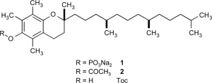
Adapted from Kato et al. 2011, Figure 1. Compound 1 is disodium tocopheryl phosphate, the TocoPhos™ active, shown with α-tocopherol and tocopheryl acetate.
IONIZATION OF THE PHOSPHATE GROUP
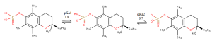
Adapted from Saleh et al. 2020, Figure 1. The phosphate undergoes two ionization steps, with pKa values of approximately 1.8 and 6.7. A predominantly dianionic state is described near pH 7.4.
CHROMANOL HEAD: ANTIOXIDANT CORE
The 6-position phenolic hydroxyl captures radicals in vitamin E; phosphorylation protects this site.
6-O-PHOSPHATE: AN IONIZABLE HANDLE
Ionization depends on pH. Near pH 7.4, the predominantly dianionic form supports water affinity while the phosphate protects the phenolic hydroxyl until hydrolysis.
SODIUM SALT: THE INCI-DEFINED FORM
Supplied as Sodium Tocopheryl Phosphate, a water-dispersible powder. The free acid has formula C29H51O5P, PubChem CID 94493.
PHYTYL TAIL: LIPID ANCHOR
After hydrolysis, the hydrophobic tail anchors released α-tocopherol in membranes, where it intercepts lipid peroxyl radicals.
Near pH 7.4, this anionic amphiphile self-associates into approximately 9 nm aggregates, with a critical aggregation concentration (CAC) of approximately 4.2 mM. At pH 9 it is monomeric, providing a formulation variable. Saleh et al. 2020.
VITAMIN E OPTIONS
Choosing the appropriate vitamin E form
Conventional forms and formulation comparison
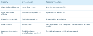
Corresponding phosphate-salt properties
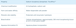
*Reconstructed human skin model: tocopherol accumulation after topical treatment, Kato et al. 2011, Figure 2A. General chemistry: Zingg 2018. Photostability of the phosphorylated form: Saleh et al. 2021.
Different systems call for different tools. The phosphate salt provides a route for aqueous and scalp-oriented formats.
UPTAKE · BIOCONVERSION · ANTIOXIDANT ACTION
Delivered as phosphate, activated as vitamin E
ACTIVATION PATHWAY
1 APPLICATION: an aqueous formulation deposits sodium tocopheryl phosphate at the skin surface.
2 UPTAKE: keratinocytes internalize the phosphorylated active.
3 BIOCONVERSION: cellular phosphatases hydrolyze the phosphate, releasing α-tocopherol.
4 ANTIOXIDANT ACTION: α-tocopherol associates with membranes and intercepts lipid peroxyl radicals.
• Deuterium tracing showed hydrolysis of α-TP to α-tocopherol in cells and mice, but not in culture medium, with lower plasma and liver lipid peroxidation. Nishio et al. 2011.
• In reconstructed human skin, tocopherol accumulation was approximately 190 nmol/g tissue at 24 hours with the phosphate salt; acetate remained near baseline. Kato et al. 2011, Figure 2A.
EVIDENCE: INTRACELLULAR BIOCONVERSION
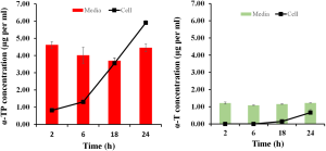
HaCaT keratinocytes were incubated with 133 µg/mL α-TP. Intracellular α-TP increased, and α-tocopherol (α-T) appeared in cell lysates at 2–24 hours, supporting uptake and enzymatic hydrolysis. Adapted from Saleh et al. 2021, Figure 8.
Saleh et al. Scientific Reports. 2021. doi:10.1038/s41598-021-01299-y
STRESS BIOLOGY
Addressing oxidative and nitrosative stress
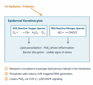
PUBLISHED EVIDENCE
UVB AND RNS IN HUMAN KERATINOCYTES
Real-time fluorescence imaging showed UVB-triggered RNS generation involving neuronal nitric oxide synthase (nNOS). Sodium dl-α-tocopheryl phosphate suppressed this response. Nakama et al. 2009.
SOOTHING-RELATED SIGNALS
In UVB-exposed NHEK, the sodium salt reduced Cox-2 mRNA by 33–51% at 0.5 µM, inhibited p38 MAPK phosphorylation and lowered PGE2. Kato et al. 2011.
MODEL CONTEXT MATTERS
Intact α-TP is a weak radical scavenger in cell-free DPPH / ORAC assays. Cellular protection follows bioconversion to α-tocopherol, making the experimental model essential to interpretation. Saleh et al. 2021, Figure 7.
KEY TERMS
ROS: reactive oxygen species. RNS: reactive nitrogen species. nNOS: neuronal nitric oxide synthase. PGE2: prostaglandin E2. NHEK: normal human epidermal keratinocytes.
EVIDENCE FOCUS: UV STRESS
Pretreated keratinocytes withstand high-dose UVA1 stress
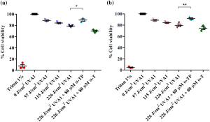
HaCaT keratinocytes were pretreated for 24 hours before UVA1 exposure. Viability was measured by Alamar Blue 24 hours later. Mean ± SD; *p < 0.05 and **p < 0.01 versus irradiated control. From Saleh et al. 2021, Figure 5.
Saleh et al. Scientific Reports. 2021. doi:10.1038/s41598-021-01299-y
VIABILITY AFTER 226 J/cm² UVA1
88% versus 70% cell viability
80 µM α-TP pretreatment versus irradiated control. α-Tocopherol at the same concentration provided no significant protection in this model.
INTRACELLULAR ROS AFTER 57 J/cm²
24.1% lower ROS versus control
100 µM α-TP; H2DCFDA measurement within 2 hours after irradiation. α-Tocopherol reduced ROS by 23.9%.
Protection coincided with phosphatase-mediated conversion to α-tocopherol. Intact α-TP was weak in cell-free assays but protective in cells. The phosphorylated form also had a higher LD50 in this model: 1676 versus 1070 µM.
Additional UVB evidence: suppression of UVB-induced RNS in human keratinocytes. Nakama et al. 2009. J SCCJ 43(1):19. doi:10.5107/sccj.43.19.
MOISTURIZATION MECHANISM
Supporting the skin’s ceramide-production machinery
Ceramides are part of the lipid mortar of the stratum corneum. In keratinocytes, sodium tocopheryl phosphate acts on upstream processes involved in their synthesis.

In NHEK, the sodium salt increased intracellular ceramides to approximately 1.8-fold at 50 µM (*), SPTLC2 expression to approximately 2.2-fold (**), differentiation markers and intracellular Ca²⁺. Kato & Takahashi 2012.
WHY THIS MATTERS TO FORMULATORS
• Support for endogenous ceramide synthesis complements formulations that directly supply ceramides.
• Differentiation markers such as TG1, involucrin and loricrin inform barrier-care positioning.
• Supporting literature reports vitamin E upregulation of transglutaminase-1 and PPARγ in human keratinocytes. De Pascale et al. 2006.
Kato & Takahashi. Bioorg Med Chem. 2012;20(12):3837–3842. PMID 22579618. The schematic synthesizes the study pathway. Fold changes are approximate readings from the original figures. SPT: serine palmitoyltransferase. TG1: transglutaminase-1. CK10: cytokeratin 10. PPARγ: peroxisome proliferator-activated receptor γ.
EVIDENCE FOCUS: MOISTURIZATION
From keratinocyte signaling to a more hydrated barrier
IN VITRO: HUMAN KERATINOCYTES, NHEK
▲ Intracellular ceramides: approximately 1.8-fold versus control at 50 µM (*).
▲ SPT genes: SPTLC2 mRNA approximately 2.2-fold (**); SPTLC1 also increased (*).
▲ Differentiation markers TGM1, KRT10, IVL and LOR: approximately 2.2–3.3-fold (*/**).
▲ Intracellular Ca²⁺: significantly higher at 50–75 µM (*), consistent with differentiation signaling.
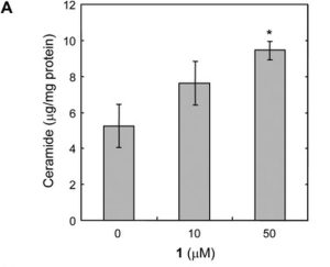
Original figure, panel 4A: intracellular NHEK ceramides increase with dose at 0 / 10 / 50 µM, measured as µg/mg protein.
IN VIVO: HAIRLESS MICE, TOPICAL 1% / 2% FOR 4 WEEKS
▲ Stratum corneum ceramides: approximately 4.8–5.0 µg/cm² in the 1% (*) and 2% (**) groups, versus approximately 3 µg/cm² in untreated and placebo groups.
▲ Hygroscopicity at 30 seconds: higher with both 1% (*) and 2% (**) versus untreated / placebo.
▲ Moisture retention at 60 seconds: significantly higher with both concentrations (*).
These findings link improved stratum corneum hydration with endogenous lipid processes.
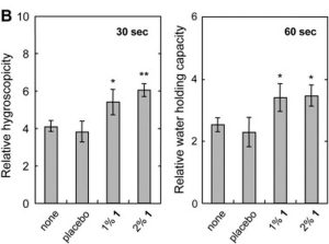
Original figure, panel 2B: stratum corneum hygroscopicity at 30 seconds and moisture retention at 60 seconds; untreated / placebo / 1% / 2%.
Kato & Takahashi. Bioorg Med Chem. 2012;20(12):3837–3842. doi:10.1016/j.bmc.2012.04.029. PMID 22579618. Panels 2B and 4A are excerpts from the paper. Fold changes are approximate readings from original axes; * / ** reproduce the authors’ significance notation.
EVIDENCE FOCUS: SKIN COMFORT
Modulating inflammatory cascades in stressed keratinocytes
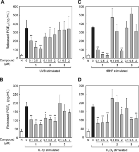
NHEK were pretreated with the sodium salt at 0.1–2 µM for 24 hours before UVB, IL-1β, tBHP or H2O2 challenge. PGE2 was measured in culture medium. Kato et al. 2011, Figure 3.
Kato et al. Bioorg Med Chem. 2011;19(21):6348–6355. doi:10.1016/j.bmc.2011.08.067
PGE2 REDUCTION VERSUS STIMULATED CONTROL
UVB: 44 / 65 / 69% at 0.1 / 0.5 / 2 µM.
tBHP: 74 / 87 / 90%; IL-1β: 41 / 58 / 57%.
H2O2: approximately 50%; UVB alone increased PGE2 approximately fivefold.
The acetate was weaker, and α-tocopherol had little effect at these concentrations.
CELL DEATH UNDER OXIDATIVE STRESS
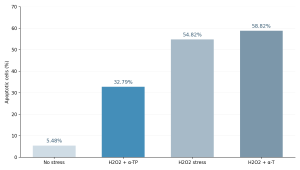
Redrawn from US20090104258A1, Example 1. Keratinocytes, 1 mM H2O2 for 24 hours; flow cytometry. The y-axis is the percentage of apoptotic cells.
Example 2 of the same patent publication: α-TP at 1 µg/mL increased large stem-cell-derived colonies by 104% in a clonogenic assay under oxidative stress.
DELIVERY SCIENCE
The vehicle influences delivery to viable skin
α-TP FLUX ACROSS A SYNTHETIC MEMBRANE
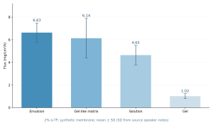
Redrawn from Saleh et al. 2023: steady-state flux of 2% α-TP from four vehicles across a synthetic membrane.
In human skin, the emulsion outperformed the gel-like system:
3× viable-layer deposition at 3 hours and 5× at 24 hours.
Human-skin flux: 328.6 versus 175.2 µg/cm²/h. Cell-free DPPH activity of the delivered dose was higher: 73% versus 46%.
SKIN DEPOSITION: pH 7.4 VERSUS α-TOCOPHEROL
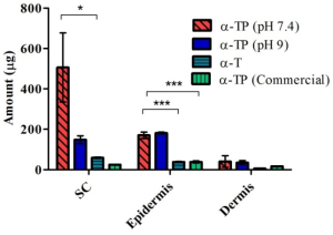
Porcine skin, 3 hours, infinite dose: α-TP at pH 7.4 reached approximately 506 ± 296 µg in the stratum corneum and 170 ± 29 µg in the epidermis. From Saleh et al. 2020, Figure 5. The source slide also describes approximately 8-fold / 4-fold differences; the comparison basis requires checking against the original paper.
At pH 7.4, α-TP forms approximately 9 nm aggregates that fuse with stratum corneum lipids in cryo-electron microscopy. At pH 9, monomer deposition is lower. Formulation pH therefore affects aggregation and skin interactions.
Saleh et al. Int J Pharm. 2023. doi:10.1016/j.ijpharm.2023.122781. PMID 36849039. Saleh et al. Int J Pharm. 2020. doi:10.1016/j.ijpharm.2020.120000.
SKINCARE APPLICATION PORTFOLIO
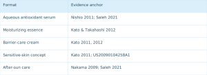
Five formats with a shared evidence base
Formats and rationale
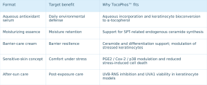
Corresponding evidence anchors
SCALPIFICATION
The scalp is skin with additional challenges
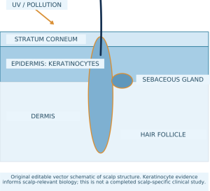
The scalp shares keratinocytes, a ceramide barrier and UV / pollution exposure with facial skin, together with dense follicles and sebum.
SHARED STRESS BIOLOGY
UV reaches the scalp surface. ROS / RNS responses documented in keratinocytes inform this application rationale. Nakama 2009; Saleh 2021.
SHARED HYDRATION MACHINERY
The ceramide barrier also matters to scalp dryness and tightness. Kato & Takahashi 2012.
SHARED COMFORT-RELATED SIGNALS
PGE2-related stress pathways provide a basis for conceptual extension to scalp comfort. Kato et al. 2011.
The scalp is a development extension for TocoPhos™. PuriPharm technical and application teams support joint scalp-formulation projects.
SCALP PROTECTION AND COMFORT
Three scalp-care opportunities
FOLLICULAR-ENVIRONMENT PROTECTION
The scalp surface is UV-exposed skin. Keratinocyte findings on UVA1 survival and UVB-triggered RNS provide a rationale for leave-on antioxidant scalp care. Nakama et al. 2009; Saleh et al. 2021.
HYDRATION BALANCE AND BARRIER SUPPORT
SPT-related ceramide synthesis and improved stratum corneum moisture retention inform concepts for dry, tight-feeling scalps. Kato & Takahashi 2012.
COMFORT FOR STRESSED SCALPS
PGE2 / Cox-2 / p38 modulation and reduced stress-induced keratinocyte death provide a rationale for comfort-oriented products. Kato et al. 2011; US20090104258A1, Example 1.
All three are development opportunities extrapolated from published epidermal evidence. Work with PuriPharm Co., Ltd. to translate them into scalp-care products.
HAIRCARE FORMATS
TocoPhos™ in scalp and haircare formulations
PRACTICAL FORMULATION
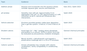
Six practical principles for phosphate formulations
SOLUBILITY VERSUS pH
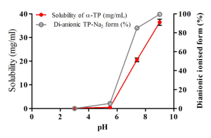
Adapted from Saleh et al. 2020, Figure 1. α-TP solubility rises with pH alongside changes in aggregation.
Contact PuriPharm technical and application teams for grade-specific use levels, processing-temperature limits, shelf life and full technical documentation.
Saleh et al. Int J Pharm. 2020. doi:10.1016/j.ijpharm.2020.120000. Saleh et al. Int J Pharm. 2023. doi:10.1016/j.ijpharm.2023.122781.
SKINCARE CONCEPT GALLERY
Three concepts to start development
FORMULATION CONCEPT
DAILY DEFENSE ANTIOXIDANT SERUM
Aqueous gel serum · Daily leave-on use
Suggested ingredients: Niacinamide · Panthenol · Glycerin
Aqueous delivery of a biologically activated antioxidant: keratinocyte uptake and conversion to α-tocopherol provide a rationale for ROS / RNS stress care.
Evidence: Nishio 2011; Saleh 2021; Nakama 2009.
FORMULATION CONCEPT
CERAMIDE-SUPPORTING MOISTURIZER
Oil-in-water cream · Daily moisturization
Suggested ingredients: Ceramide NP · Glycerin · Squalane
Two complementary routes: direct ceramide supplementation and support for endogenous SPT-related synthesis.
Evidence: Kato & Takahashi 2012.
FORMULATION CONCEPT
AFTER-SUN CARE LOTION
Light emulsion · After-sun comfort
Suggested ingredients: Sodium Ascorbyl Phosphate · Bisabolol · Betaine
A reported combination with magnesium ascorbyl phosphate inhibited PGE2 more than either ingredient alone. This informs a vitamin C phosphate combination concept; the proposed sodium derivative is not the identical tested combination.
Evidence: Kato 2011, Figure 6; Nakama 2009.
These concepts have published mechanistic evidence anchors. Contact PuriPharm Co., Ltd. for formulation collaboration.
SCALP AND HAIRCARE CONCEPT GALLERY
Scalp-first formats informed by skin science
FORMULATION CONCEPT
ANTIOXIDANT SCALP SERUM
Aqueous leave-on serum · Daily use
Suggested ingredients: Niacinamide · Panthenol · Glycerin
A scalp-surface serum with biologically activated antioxidant positioning, derived from epidermal UV-stress evidence.
Evidence: Nakama 2009; Saleh 2021.
FORMULATION CONCEPT
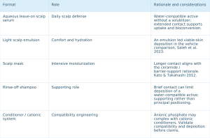
LIGHT SCALP COMFORT EMULSION
Fluid emulsion · Leave-on comfort
Suggested ingredients: Bisabolol · Betaine · Glycerin
Emulsion vehicles provided greater viable-skin deposition in the cited comparison. PGE2 modulation informs a scalp-comfort concept.
Evidence: Saleh 2023; Kato 2011.
FORMULATION CONCEPT
MOISTURIZING SCALP MASK
Cream mask · Weekly intensive care
Suggested ingredients: Ceramide NP · Butyrospermum Parkii (Shea) Butter
Longer contact is consistent with a ceramide-synthesis rationale for dry, tight-feeling scalps.
Evidence: Kato & Takahashi 2012.
Develop a coordinated scalp-care range, from serums to masks, with PuriPharm Co., Ltd.
FOUR KEY POINTS
Why PuriActives® TocoPhos™?
PROTECTED AND BIOLOGICALLY AVAILABLE
Phosphate chemistry protects the antioxidant site, with cellular phosphatases releasing α-tocopherol after uptake.
EVIDENCE FROM RELEVANT MODELS
Human keratinocytes, reconstructed skin, in vivo barrier models and human skin delivery studies provide traceable evidence.
FORMULATION DEVELOPMENT SUPPORT
Aqueous-phase use, published vehicle and pH data, and preservative-testing evidence from an emulsion prototype.
ONE ACTIVE, TWO CARE AREAS
Skin defense and moisturization, alongside scalp-care concepts extrapolated from the same evidence base.
Vitamin E, reimagined for formulation development.
Work with us on your next skincare or scalp-care concept.
PuriPharm Co., Ltd. · Technical and Application Team
SCIENTIFIC REFERENCES
Scientific references
1. Nakama M. et al. Inhibitory effect of sodium dl-α-tocopheryl-6-O-phosphate on UVB-induced reactive nitrogen species in human keratinocytes. J Soc Cosmet Chem Jpn 43(1):19–25, 2009. doi:10.5107/sccj.43.19
2. Kato E., Sasaki Y., Takahashi N. Sodium dl-α-tocopheryl-6-O-phosphate inhibits PGE2 production in keratinocytes induced by UVB, IL-1β and peroxidants. Bioorg Med Chem 19(21):6348–6355, 2011. doi:10.1016/j.bmc.2011.08.067. PMID 21945458.
3. Kato E., Takahashi N. Improvement by sodium dl-α-tocopheryl-6-O-phosphate treatment of moisture-retaining ability in stratum corneum through increased ceramide levels. Bioorg Med Chem 20(12):3837–3842, 2012. doi:10.1016/j.bmc.2012.04.029. PMID 22579618.
4. Nishio K. et al. α-Tocopheryl phosphate is hydrolyzed to α-tocopherol in cells and in vivo. Free Radic Biol Med 50(12):1794–1800, 2011. PMID 21443945.
5. Saleh M. et al. Tocopheryl phosphate in keratinocyte UV-stress models. Scientific Reports 11:22400, 2021. doi:10.1038/s41598-021-01299-y
6. Gianello R. et al. α-Tocopheryl phosphate: chemistry and biological activity. Free Radic Biol Med 39(7):970–976, 2005. PMID 16140216.
7. Nakayama S. et al. Protective effect of α-tocopheryl phosphate against UVB-induced damage in cultured mouse skin. J Invest Dermatol 121:406–411, 2003.
8. De Pascale M.C. et al. Vitamin E upregulates transglutaminase-1 and PPARγ in human keratinocytes. Arch Biochem Biophys 447:97–106, 2006.
9. Zingg J.-M. Vitamin E: regulatory role beyond antioxidant activity. Adv Food Nutr Res 83:311–363, 2018.
10. Ghayour-Mobarhan M. et al. Tocopheryl phosphate: a review of biological properties. J Diet Suppl, 2014. doi:10.3109/19390211.2014.952859
11. Ogru E. et al. Vitamin E phosphate: an endogenous form of vitamin E. Free Radic Res, 2003.
FORMULATION AND PATENT REFERENCES
Formulation and patent references
12. Saleh M. et al. Ionization, pH-dependent solubility, self-association and skin deposition of α-tocopheryl phosphate. Int J Pharm 594:120000, 2020. doi:10.1016/j.ijpharm.2020.120000
13. Saleh M. et al. Vehicle-dependent delivery of α-tocopheryl phosphate to viable skin. Int J Pharm 636:122781, 2023. PMID 36849039 · doi:10.1016/j.ijpharm.2023.122781
14. Patent publication US20090104258A1 (published 2009), cited by publication number and examples: Example 1 (oxidative-stress-induced keratinocyte apoptosis, flow cytometry) and Example 2 (clonogenic assay; large stem-cell-derived colonies +104%).
15. Gavin A. et al. Tocopheryl phosphate mixture (TPM) as a skin-delivery enhancer for co-formulated actives. Drug Deliv Transl Res, 2016. doi:10.1007/s13346-016-0331-x
16. Mozafari M. et al. Tocopheryl phosphate-based delivery systems (review). Int J Transl Res.
NanoActive EGCG 纳米脂质体
NanoActive EGCGLiposomal Epigallocatechin Gallate
PRODUCT & BRAND
NanoActive EGCG · Nanoliposomes

Powered by PURISOME® FlexVes™ Liposome Technology
PuriPharm Co., Ltd. · NanoActive platform ingredient
PRODUCT PLATFORM
One catechin platform, multiple formulation opportunities
EGCG is one of the most studied polyphenols in skin biology, but its practical formulation value depends on protection and delivery.
NanoActive EGCG combines this multifunctional active with liposomal delivery across five evidence-informed skin and scalp development areas.
SKIN
ANTIOXIDANT DEFENSE
Resilience under UV and urban environmental stress.
PHOTOAGING-RELATED MATRIX PROTECTION
Support for collagen-matrix signaling and youthful skin texture.
SEBUM BALANCE AND BLEMISH-PRONE SKIN
Sebocyte lipid biology supported by clinical lesion-count evidence.
A MORE EVEN-TONED APPEARANCE
Melanogenesis signaling rationale from cell studies.
SCALP AND FOLLICLES
SCALP COMFORT AND FOLLICULAR VITALITY
Evidence from dermal papilla cells and human follicle organ culture.
Each development area is based on the scientific literature cited in its corresponding section.
KEY ACTIVE
The active behind the platform
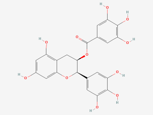
Epigallocatechin gallate (EGCG): 2D structure, PubChem CID 65064.
ACTIVE INCI
EPIGALLOCATECHIN GALLATE
MOLECULAR FORMULA: C22H18O11 · MOLAR MASS: 458.37 g/mol
BOTANICAL SOURCE: the principal catechin in green tea (Camellia sinensis), formed by esterification of a flavan-3-ol with gallic acid.
STRUCTURE–ACTIVITY RELATIONSHIP: eight phenolic hydroxyl groups support radical scavenging but also confer oxidation sensitivity.
PRODUCT POSITIONING
NanoActive EGCG presents the catechin in a liposomal format built on PuriPharm’s PURISOME® platform to protect the active and support delivery to skin.
The INCI shown identifies the active ingredient. Refer to product technical documentation for the complete composition of the liposomal dispersion.
Structure source: PubChem CID 65064, NIH National Library of Medicine.
FORMULATION RATIONALE
Formulation determines EGCG performance
EGCG IN SOLUTION
DEGRADATION DRIVERS
Oxygen and light
Higher temperature
Neutral to alkaline pH
Vehicle and ionic environment
Oxidation and degradation of catechins in solution
IMPLICATIONS FOR FORMULATORS
Protect the active from oxygen, light and heat during processing and storage.
Design the vehicle and pH carefully.
Consider encapsulation to protect the catechin before use.
Design a delivery route to relevant skin compartments.
EGCG is valuable but chemically vulnerable. Solution studies show that pH, temperature and vehicle affect how much intact EGCG remains available, providing the rationale for protection and delivery engineering.
Proniuk S, Liederer BM, Blanchard J. J Pharm Sci. 2002;91(1):111–116. DOI: 10.1002/jps.10009. PMID: 11782902.
DELIVERY TECHNOLOGY
Powered by PURISOME® FlexVes™ Liposome Technology
EGCG
Skin-compatible phospholipid bilayer vesicles
Schematic, not to scale. Refer to PuriPharm technical documentation for the actual structure.
1 PROTECT VULNERABLE ACTIVES
Protect oxidation- and degradation-sensitive ingredients such as EGCG to support shelf-life quality.
2 SUPPORT LOCAL SKIN DELIVERY
Enhance stratum corneum penetration and the bioavailability of encapsulated actives.
3 SUSTAINED AND CONTROLLED RELEASE
Extend release, with a design rationale for reducing irritation risk.
4 FORMULATION COMPATIBILITY
Support compatibility and skin feel in leave-on and rinse-off systems.
All NanoActive products use the PURISOME® platform, drawing on PuriPharm’s more than ten years of nanodelivery R&D and industrial production experience.
PURISOME® platform source: PuriPharm Co., Ltd. brand documentation, V1.0 (2026).
DELIVERY RATIONALE: PUBLISHED RESEARCH
Delivery research informs formulation design
PUBLISHED FIGURE: SKIN DEPOSITION OF EGCG (µg/cm²)
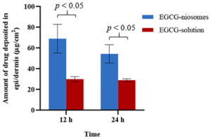
Reproduced from Li et al. 2022, Figure 5 (CC BY 4.0). Mean ± SD, n = 3. EGCG-loaded niosomes are surfactant vesicles. This study informs delivery design and did not measure PURISOME® performance.
PUBLISHED FIGURE: CUMULATIVE EGCG RELEASE (%)
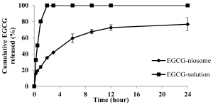
Compared with a simple solution, vesicular encapsulation approximately doubled EGCG deposition in human skin and provided sustained release over 24 hours.
Vesicle engineering can support surface residence, time-dependent release and delivery into viable skin. These findings inform the design rationale for the PURISOME® liposome platform.
Li D et al. Pharmaceutics. 2022;14(4):726. DOI: 10.3390/pharmaceutics14040726. PMID: 35456560.
ADDITIONAL DATA FROM THE SOURCE SPEAKER NOTES
Ex vivo human skin deposition at 12 hours was 69.0 ± 13.87 µg/cm² with niosomes versus 30.02 ± 2.45 µg/cm² with solution. At 24 hours, values were 54.38 ± 8.86 versus 29.00 ± 1.36 µg/cm² (mean ± SD, n = 3). Niosomes showed biphasic release, approximately 35% at 3 hours and 73% at 24 hours; Korsmeyer–Peppas fit r² = 0.996. These are Li et al.’s niosome results, not PURISOME® measurements.
MECHANISM OVERVIEW
From molecular activity to skin appearance
EGCG
One active, five pathways
INFLAMMATORY SIGNALING
Sebocytes and PM10-exposed keratinocytes. Yoon 2013; Seok 2018.
REDOX DEFENSE
Human UV-challenged skin and oxidative endpoints in fibroblasts. Katiyar 2001; Jia 2023.
SEBOCYTE LIPID BIOLOGY
AMPK / SREBP-1 in human sebocytes. Yoon 2013.
MATRIX PROTECTION
MMP-1/3/10, TIMP-1 and TGF-β1 expression. Jia 2023.
MELANOGENESIS SIGNALING
cAMP / CREB / MITF in B16F10 cells. Zhang 2020.
These experimentally studied pathways inform formulation concepts for resilience, clarity, even tone and follicular vitality.
Pathway overview based on Katiyar 2001; Yoon 2013; Seok 2018; Zhang 2020; Jia 2023. Full references appear at the end.
SKIN SCIENCE: ANTIOXIDANT DEFENSE
Antioxidant evidence in human skin
HYDROGEN PEROXIDE GENERATION IN UV-CHALLENGED SKIN
−68%
−90%
0 25 50 75 100 — Reduction versus UV-only control (%)
EPIDERMAL LIPID PEROXIDATION
−41%
−84%
0 25 50 75 100 — Reduction versus UV-only control (%)
STUDY CONTEXT
EGCG was applied topically to human skin before a single UV challenge of four minimal erythema doses. The ranges cover post-exposure assessment time points. EGCG also helped preserve glutathione-related antioxidant defenses.
These are biological stress-defense endpoints, not SPF values or evidence of UV filtering.
FORMULATION RATIONALE
UV exposure can generate ROS faster than endogenous defenses can clear them.
The reduced oxidative burden observed in human skin supports antioxidant and resilience-oriented daily-care concepts.
Katiyar SK et al. Carcinogenesis. 2001;22(2):287–294. DOI: 10.1093/carcin/22.2.287. PMID: 11181450.
SKIN SCIENCE: PHOTOAGING
Matrix support under photoaging stress
PUBLISHED FIGURE: MATRIX MARKERS IN UVA-STRESSED FIBROBLASTS
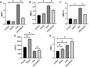
Reproduced from Jia et al. 2023, Figure 6, panels A–E. Human dermal fibroblasts, UVA model, EGCG 25 µg/mL.
REPORTED FINDINGS
MMP-1 / MMP-3 / MMP-10 expression decreased versus UVA alone (panels A–C).
TGF-β1 secretion increased (panel D; matrix-building signal).
TIMP-1 mRNA increased (panel E; metalloproteinase inhibitor).
Degradation-associated signals decreased as matrix-supporting signals recovered.
ENDPOINTS
MMP-1/3/10 and TIMP-1 mRNA were measured by RT-PCR. Secreted TGF-β1 protein was measured in culture supernatants (*p < 0.05).
Photoaging involves an imbalance between matrix degradation and renewal. EGCG modulated this balance in cultured human fibroblasts, providing a mechanistic basis for photoaging-oriented formulations.
Jia Y et al. Clin Cosmet Investig Dermatol. 2023;16. DOI: 10.2147/CCID.S398547. PMID: 36704608.
SKIN SCIENCE: CELLULAR SENESCENCE
Reducing the cellular burden of UVA stress
Repeated UVA exposure can promote cellular senescence, with loss of cell division and altered tissue signaling.
Senescence-associated β-galactosidase (SA-β-gal) staining identifies blue-stained cells, quantified as a percentage.
In human fibroblasts, EGCG nearly halved the senescent-cell proportion after UVA exposure.
MODEL
Human dermal fibroblasts; EGCG 25 µg/mL for 24 hours. Mean ± SD from three independent experiments (*p < 0.05).
PUBLISHED FIGURE: SA-β-GAL STAINING AND QUANTIFICATION
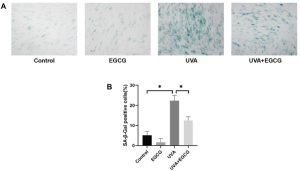
Approximately 45% lower senescent-cell proportion relative to UVA alone.
Reproduced from Jia et al. 2023, Figure 2. (A) SA-β-gal staining. (B) Quantification: 23.07% with UVA versus 12.75% with UVA + EGCG.
Jia Y et al. Clin Cosmet Investig Dermatol. 2023;16. DOI: 10.2147/CCID.S398547. PMID: 36704608.
SKIN SCIENCE: URBAN ENVIRONMENTAL STRESS
A scientific rationale for urban skin care
AIRBORNE PARTICULATE MATTER: PM10
Particles that contact facial skin and the scalp during everyday exposure.
KERATINOCYTE STRESS RESPONSE
Oxidative stress and inflammatory signaling increase in exposed cells.
EGCG AT 3–10 µM
Attenuation of oxidative and inflammatory responses in PM10-exposed cells.
Urban exposure places an ongoing particulate burden on skin and scalp. Micromolar EGCG attenuated exposure-related stress in primary human keratinocytes.
These findings support a mechanistic rationale for antioxidant day creams, serums and leave-on scalp products focused on environmental resilience.
MODEL
Primary human keratinocytes exposed to PM10; EGCG tested at 3–10 µM.
Mechanistic evidence only: the study did not measure a pollution-blocking percentage for a finished product.
Seok JK et al. Skin Pharmacol Physiol. 2018;31(3):134–143. DOI: 10.1159/000487400. PMID: 29566388.
SKIN SCIENCE: TONE
Supporting a more even-toned appearance
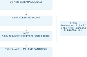
PUBLISHED FIGURES: ZHANG ET AL. 2020, B16F10 CELLS
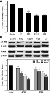
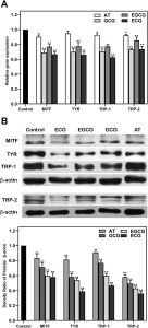
Figure 4: cellular tyrosinase activity (A) and melanin content (B). Figure 5: MITF / TYR / TRP-1 / TRP-2 proteins (B) and cAMP (C).
Melanogenesis follows a signaling cascade: increased cAMP, CREB activation and MITF-driven tyrosinase expression and melanin synthesis. EGCG modulated this cascade in B16F10 cells, supporting a mechanistic rationale for even-tone and targeted-care concepts. These are cell-level findings, not clinical age-spot results.
CELL CONTEXT
B16F10 melanocyte model. The schematic presents the pathway, while the published figures show experimental data.
Zhang X et al. Fitoterapia. 2020;145:104634. DOI: 10.1016/j.fitote.2020.104634. PMID: 32454171.
SKIN SCIENCE: SEBUM BIOLOGY
Addressing excess lipid production at the cellular level

AMPK: AMP-activated protein kinase, a cellular energy sensor that suppresses anabolic processes.
SREBP-1: sterol regulatory element-binding protein 1, a transcription factor that activates sebocyte lipid-synthesis genes.
Sebum synthesis in sebocytes is regulated by SREBP-1. In cultured human sebocytes, EGCG activated AMPK and reduced this lipogenic program.
This provides cellular evidence relevant to oil-balancing and blemish-prone-skin concepts.
MODEL
Cultured human sebocytes. Clinical evidence from the same study is presented below.
PUBLISHED FIGURE: YOON ET AL. 2013, FIGURE 1
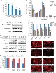
Sebum, AMPK and SREBP-1 measurements in human sebocytes.
Yoon JY et al. J Invest Dermatol. 2013;133(2):429–440. DOI: 10.1038/jid.2012.292. PMID: 23096708.
SKIN SCIENCE: CLINICAL EVIDENCE
Clinical evidence in blemish-prone skin
−89%
Reported reduction in inflammatory lesion count versus baseline in the 1% EGCG group.
PUBLISHED FIGURES: YOON ET AL. 2013, SPLIT-FACE CONTROLLED TRIAL
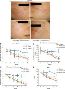
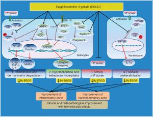
Figure 4 presents clinical photographs, Leeds scores and lesion-count changes. The other supplied image is a mechanistic summary schematic, rather than a split-face photograph.
STUDY DESIGN
Split-face, vehicle-controlled study; the contralateral side received vehicle.
1% or 5% EGCG, twice daily for 8 weeks.
35 of 37 participants completed: 17 in the 1% group and 18 in the 5% group.
Endpoints: inflammatory and non-inflammatory lesion counts.
This controlled human study connects sebocyte biology with visible changes in blemish-prone skin, providing a clinical anchor within the evidence base.
Yoon JY et al. J Invest Dermatol. 2013;133(2):429–440. DOI: 10.1038/jid.2012.292. PMID: 23096708.
SKIN SCIENCE: SEBUM, MICROBIAL INTERACTIONS AND COMFORT
Linking sebum, microbial interactions and skin comfort
1 · LIPID ENVIRONMENT
Excess sebum contributes to blemish-prone conditions.
2 · MICROBIAL INTERACTIONS
Cutibacterium acnes proliferates in lipid-rich follicles.
3 · INFLAMMATION AND DISCOMFORT
Inflammatory signaling contributes to visible blemishes.
EXPERIMENTALLY STUDIED EGCG EFFECTS
GROWTH INHIBITION
EGCG showed in vitro inhibition of C. acnes, tested under its historical name Propionibacterium acnes ATCC 6919.
INFLAMMATORY SIGNALING
EGCG attenuated NF-κB-related responses in human sebocytes.
Together with effects on sebocyte lipids, these findings provide a multi-level rationale for blemish-care formulation: less lipid substrate, measured microbial inhibition and reduced inflammatory signaling.
PUBLISHED FIGURE: YOON 2013, FIGURE 2
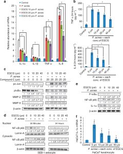
Sebocyte NF-κB / AP-1 inflammatory measurements.
Yoon JY et al. J Invest Dermatol. 2013;133:429–440. Widyaningrum N et al. Asian J Biol Sci. 2015;8(2):93–101. DOI: 10.3923/ajbs.2015.93.101.
APPLICATION CONCEPTS: SKIN
Skin application concepts
Targets and scientific rationale
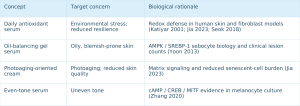
Formats and formulation focus
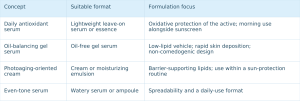
These are formulation-development concepts, not tested finished products. Confirm performance at the product level.
Evidence sources are cited in the skin-science sections; full references appear at the end.
SCALP AND FOLLICLE SCIENCE
Bringing skincare science to scalp care
The scalp faces oxidative and inflammatory challenges similar to facial skin, alongside a distinctive structure: the follicle, a living mini-organ that produces hair.
SCALP ENVIRONMENT
Surface skin exposed to UV, pollution and sebum oxidation.
FOLLICLE
Living cells that drive the hair cycle, including dermal papilla signaling cells.
HAIR SHAFT
A non-living fiber whose surface can be conditioned but whose structure is produced by the follicle.
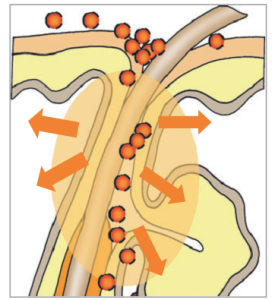
Scalp and follicle anatomy: conceptual illustration from PuriPharm’s asset collection.
Anatomical schematic for structural reference only.
SCALP AND FOLLICLE SCIENCE
A rationale for a more resilient scalp environment
EVERYDAY SCALP STRESSORS
UV, airborne particles, sebum oxidation and styling stress also challenge the scalp.
OXIDATIVE AND INFLAMMATORY LOAD
Redox and inflammatory pathways documented in skin are relevant to perifollicular scalp tissue.
EGCG DEVELOPMENT RATIONALE
Antioxidant defense and inflammatory modulation formulated for the scalp surface.
A resilient scalp surface provides a basis for follicle-focused care.
NanoActive EGCG brings documented redox and inflammatory biology to leave-on scalp concepts, where longer contact supports local availability.
This is a mechanistic extension from skin studies to scalp-care development. Specific cell and follicle evidence follows.
Rationale based on Katiyar 2001; Seok 2018; Jia 2023. Full references appear at the end.
SCALP AND FOLLICLE SCIENCE: DERMAL PAPILLA
Activity in human dermal papilla cells
Dermal papilla cells (DPCs), located at the follicle base, are signaling cells involved in the hair cycle.
In cultured human DPCs, EGCG at 0.01–0.5 µM produced a significant, dose-dependent proliferative response by MTT assay (p < 0.05).
These micromolar responses provide a cellular basis for follicular-vitality concepts.
EXPERIMENT
MTT proliferation assay in cultured human DPCs; Kwon et al. 2007, Figure 1. This is a cellular response, not a clinical hair-density measurement.
PUBLISHED FIGURE: KWON ET AL. 2007, FIGURE 1, MTT ASSAY
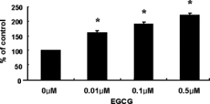
DPC proliferation increased with dose versus control at 0.01–0.5 µM (*p < 0.05). The source figure has low resolution.
Kwon OS et al. Phytomedicine. 2007;14(7–8):551–555. DOI: 10.1016/j.phymed.2006.09.009. PMID: 17092697.
SCALP AND FOLLICLE SCIENCE: SIGNALING
Signals associated with follicular vitality
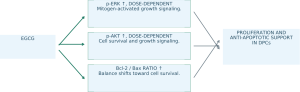
EGCG
p-ERK ↑, DOSE-DEPENDENT
Mitogen-activated growth signaling.
p-AKT ↑, DOSE-DEPENDENT
Cell survival and growth signaling.
Bcl-2 / Bax RATIO ↑
Balance shifts toward cell survival.
PROLIFERATION AND ANTI-APOPTOTIC SUPPORT IN DPCs
INTERPRETING THE RESULTS
Directional summary of Kwon et al. 2007, Figures 2–4, in cultured human DPCs.
This differs from the sebocyte apoptosis reported with EGCG: different cell types can have different outcomes.
PUBLISHED FIGURES: KWON ET AL. 2007, FIGURES 2–4
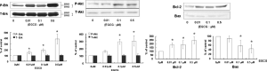
Responses of p-ERK, p-AKT and Bcl-2/Bax to EGCG in human DPCs. The source figures have low resolution.
EGCG modulated growth (ERK), survival (Akt) and apoptotic balance (Bcl-2/Bax) signals, providing complementary mechanisms relevant to follicular formulation concepts.
Kwon OS et al. Phytomedicine. 2007;14(7–8):551–555. DOI: 10.1016/j.phymed.2006.09.009. PMID: 17092697.
SCALP AND FOLLICLE SCIENCE: ORGAN CULTURE
Evidence from human follicle organ culture
FOLLICLE ELONGATION AFTER 10 DAYS, AS A PERCENTAGE OF VEHICLE CONTROL
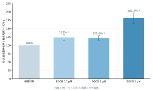
Redrawn from Kwon et al. 2007. Mean ± SD: the three EGCG groups had SDs of 9.0, 7.1 and 15.8 percentage points, respectively; p < 0.05 versus vehicle. Human follicles came from three donors and were cultured for 10 days.
Beyond isolated cells, EGCG increased elongation of whole human follicles over 10 days in organ culture.
This model bridges cellular experiments and clinical research and provides preclinical support for follicular-vitality concepts in leave-on scalp care.
EX VIVO HUMAN FOLLICLE MODEL
The same paper also reports a separate, small scalp-biomarker experiment, summarized in the source speaker notes. Neither constitutes a clinical hair-growth outcome.
Kwon OS et al. Phytomedicine. 2007;14(7–8):551–555. DOI: 10.1016/j.phymed.2006.09.009. PMID: 17092697.
ADDITIONAL CONTEXT FROM THE SOURCE SPEAKER NOTES
The paper separately reports a 4-day scalp-biomarker experiment in three volunteers using 10% EGCG in ethanol, with increased dermal papilla p-Erk/p-Akt and Bcl-2/Bax. This small experiment is distinct from ex vivo follicle elongation, is not a clinical hair-growth outcome and is not a product use-level recommendation.
SCALP AND FOLLICLE SCIENCE: DELIVERY DESIGN
Designing local skin and follicular delivery
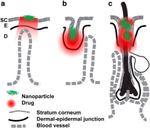
Conceptual schematic of follicular accumulation of nanocarriers, from PuriPharm’s asset collection.
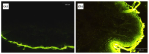
Published figure: Li et al. 2022, Figure 6. FITC-loaded vesicles concentrated at follicular openings in ex vivo human skin.
This delivery-design concept is consistent with published vesicle research (Li 2022). The image does not establish transport of intact vesicles to the dermal papilla, and smaller particle size does not automatically mean deeper penetration.
Delivery reference: Li D et al. Pharmaceutics. 2022;14(4):726. DOI: 10.3390/pharmaceutics14040726.
APPLICATION CONCEPTS: HAIR AND SCALP
Hair and scalp formulation concepts
Targets and formulation focus
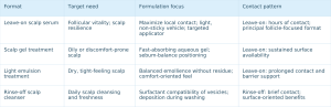
Formats and contact patterns
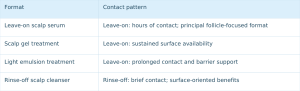
These are development concepts. Follicle-focused positioning centers on leave-on formats, where contact duration supports local activity.
Evidence sources are cited in the scalp and follicle sections. Full references appear at the end.
FORMULATION DESIGN
Targeted active combinations
NanoActive EGCG provides the core active, while complementary ingredients refine the concept. These are formulation choices; confirm compatibility in the final vehicle.
+ NIACINAMIDE
OIL-BALANCING CONCEPT
EGCG addresses sebocyte lipid biology, while niacinamide adds barrier and tone support. Complementary pathways inform oily- and blemish-prone-skin positioning.
+ PANTHENOL
COMFORT AND SOOTHING CONCEPT
EGCG modulates stress signaling, while panthenol supports hydration and comfort. This pairing is proposed for sensitive-skin and scalp formats.
+ CAFFEINE
SCALP-VITALITY CONCEPT
EGCG dermal papilla biology complements the established use of caffeine in leave-on scalp tonics and serums.
Check vehicle pH, ionic environment, oxidation drivers and preservation together. Factors governing EGCG stability (Proniuk 2002) also apply to multi-active formulations.
Combination concepts: PuriPharm. Stability principles: Proniuk 2002, discussed in the formulation-rationale section.
FORMULATION PRACTICE
Practical formulation integration
ADDITION AND HANDLING
Evaluate cool-down addition to reduce heat exposure.
Review shear exposure during homogenization and pumping.
Minimize residence at high temperature.
BASE ENVIRONMENT
Set pH deliberately; EGCG is more stable in acidic conditions.
Control ionic conditions that may challenge vesicle integrity.
Manage dissolved oxygen, light and pro-oxidant metals.
COMPATIBILITY
Screen surfactants for vesicle compatibility; high levels may disrupt lipid vesicles.
Check interactions with other actives, electrolytes and thickeners.
Confirm preservative-system compatibility.
These are development considerations, not validated processing limits. Refer to PuriPharm’s NanoActive EGCG technical documentation for product-specific addition stage, temperature and use-level guidance.
Stability principles: Proniuk 2002. Product instructions follow PuriPharm technical documentation.
FORMULATION PRACTICE
Protecting active quality through formulation and packaging
FORMULATION
Liposomal encapsulation protects EGCG from bulk-phase oxidation.
Design pH and antioxidant support around catechin chemistry.
Assess metal interactions and use chelation where appropriate.
PACKAGING
Limit light using opaque or UV-protective packaging.
Limit oxygen ingress using airless or low-headspace formats.
Match the pack to application and dose control.
MONITORING
Track color and EGCG content during stability studies.
Characterize carrier integrity, vesicle structure and size.
Use sensory changes as early-warning signals.
Active quality is a system property involving chemistry, packaging and monitoring. Finished-product shelf life and stability claims require product-level testing.
Framework based on EGCG solution chemistry (Proniuk 2002) and vesicular delivery research (Li 2022).
FROM SCIENCE TO BRAND
Translating science into brand value
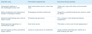
Consumer wording is limited to cosmetic positioning and anchored to the cited skin and follicle studies.
Evidence anchors: Katiyar 2001; Jia 2023; Yoon 2013; Zhang 2020; Kwon 2007.
CONCLUSION
NanoActive EGCG: from catechin science to formulation opportunity
1 MULTIFUNCTIONAL ACTIVE
Published biological evidence covers antioxidant defense, photoaging, sebum balance, tone and follicular vitality.
2 PURPOSEFUL DELIVERY
PURISOME® liposome technology is designed to protect the catechin and support local, sustained delivery.
3 SKIN AND SCALP OPPORTUNITIES
One platform spans serums, gels, moisturizers, essences, treatments and scalp-care products.
Powered by PURISOME® FlexVes™ Liposome Technology
PuriPharm Co., Ltd.
www.puriactives.com
REFERENCES · SKIN BIOLOGY
Key scientific references: skin biology
Katiyar SK et al. Green tea polyphenol (−)-epigallocatechin-3-gallate treatment of human skin inhibits UV-induced oxidative stress. Carcinogenesis. 2001;22(2):287–294. DOI: 10.1093/carcin/22.2.287. PMID: 11181450.
Jia Y et al. (−)-Epigallocatechin-3-gallate protects human skin fibroblasts from ultraviolet A induced photoaging. Clinical, Cosmetic and Investigational Dermatology. 2023;16. DOI: 10.2147/CCID.S398547. PMID: 36704608. PMCID: PMC9871046.
Seok JK, Lee JW, Kim YM, Boo YC. Punicalagin and (−)-epigallocatechin-3-gallate rescue cell viability and attenuate inflammatory responses of human epidermal keratinocytes exposed to airborne particulate matter PM10. Skin Pharmacology and Physiology. 2018;31(3):134–143. DOI: 10.1159/000487400. PMID: 29566388.
Yoon JY et al. Epigallocatechin-3-gallate improves acne in humans by modulating intracellular molecular targets and inhibiting P. acnes. Journal of Investigative Dermatology. 2013;133(2):429–440. DOI: 10.1038/jid.2012.292. PMID: 23096708.
Zhang X et al. Anti-melanogenic effects of epigallocatechin-3-gallate (EGCG), epicatechin-3-gallate (ECG) and gallocatechin-3-gallate (GCG) via down-regulation of cAMP/CREB/MITF signaling pathway in B16F10 melanoma cells. Fitoterapia. 2020;145:104634. DOI: 10.1016/j.fitote.2020.104634. PMID: 32454171.
Widyaningrum N, Fudholi A, Sudarsono, Setyowati EP. Stability of epigallocatechin gallate (EGCG) from green tea (Camellia sinensis) and its antibacterial activity against Staphylococcus epidermidis ATCC 35984 and Propionibacterium acnes ATCC 6919. Asian Journal of Biological Sciences. 2015;8(2):93–101. DOI: 10.3923/ajbs.2015.93.101.
Only literature cited in the presentation is listed. Microbial names retain the historical terminology used at publication.
Skin biology references. Hair, delivery and formulation references follow.
REFERENCES · HAIR, DELIVERY AND FORMULATION
Key scientific references: hair, delivery and formulation
HAIR AND SCALP
Kwon OS, Han JH, Yoo HG, Chung JH, Cho KH, Eun HC, Kim KH. Human hair growth enhancement in vitro by green tea epigallocatechin-3-gallate (EGCG). Phytomedicine. 2007;14(7–8):551–555. DOI: 10.1016/j.phymed.2006.09.009. PMID: 17092697.
DELIVERY AND FORMULATION
Li D et al. Niosomal nanocarriers for enhanced dermal delivery of epigallocatechin gallate for protection against oxidative stress of the skin. Pharmaceutics. 2022;14(4):726. DOI: 10.3390/pharmaceutics14040726. PMID: 35456560. PMCID: PMC9029719.
Proniuk S, Liederer BM, Blanchard J. Preformulation study of epigallocatechin gallate, a promising antioxidant for topical skin care products. Journal of Pharmaceutical Sciences. 2002;91(1):111–116. DOI: 10.1002/jps.10009. PMID: 11782902.
Structure reference: Epigallocatechin gallate, PubChem CID 65064, NIH National Library of Medicine (active-identity section).
Only literature cited in the presentation is listed. PURISOME® platform descriptions are based on PuriPharm Co., Ltd. brand documentation.
NanoActive EGCG nanoliposomes: scientific marketing report. PuriPharm Co., Ltd.
PuriActives® Crystal Retinal
PuriActives® Crystal RetinalCyclodextrin-Included Retinal
PuriPharm Co., Ltd. · Retinal science
PuriActives® Crystal Retinal Cyclodextrin-Included Retinal
High-performance retinal through molecular inclusion science
Retinal · Cyclodextrin inclusion · Skin and scalp renewal
Scientific and technical report for cosmetic formulators and brand R&D teams
PART I · NEXT-GENERATION RETINAL
The evolution of vitamin A skincare
RETINYL ESTERS
Storage forms requiring hydrolysis before entering metabolism.
RETINOL
An established cosmetic benchmark, oxidized to retinal in skin.
RETINAL (RAL)
One oxidation step from retinoic acid: its direct metabolic precursor.
RETINOIC ACID
The receptor-active form; prescription-only in many markets.
Esterases · Retinol dehydrogenases (RDH/ADH) · Retinaldehyde dehydrogenases (ALDH1A)
FROM STORAGE FORM TO SIGNALING MOLECULE
Cosmetic vitamin A ingredients follow the skin’s own metabolic pathway. Moving closer to retinoic acid reduces the number of enzymatic conversions.
Retinal is the direct biosynthetic precursor of retinoic acid, the active form that binds nuclear retinoid receptors and drives epidermal renewal.
Metabolic proximity is a mechanistic rationale, not a clinical conclusion. Relative efficacy requires controlled human studies, reviewed in Part III.
2 → 1
Oxidation steps to retinoic acid: two for retinol, one for retinal.
1994
Topical retinal was first biologically characterized in human skin (Saurat et al.).
125+
Participants indicated in the source’s milestone vehicle-controlled profilometry trial comparing retinal with retinoic acid (1998).
Saurat JH et al. J Invest Dermatol. 1994;103(6):770–774. Creidi P et al. J Am Acad Dermatol. 1998;39(6):960–965. Retinoid metabolism: RDH/ADH and ALDH1A enzyme families.
PART I · NEXT-GENERATION RETINAL
One conversion from retinoic acid: retinal biology
All-trans-retinal (C20H28O; molecular weight 284.4)
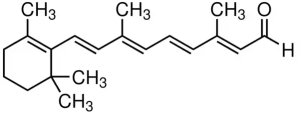
The β-ionone ring, conjugated polyene chain and terminal aldehyde underlie both biological activity and chemical vulnerability.
The conjugated system responsible for sensitivity to light and oxidation also contributes to retinal’s biological activity.
Stabilizing this molecule without changing its chemistry is the formulation challenge addressed by cyclodextrin inclusion.
A NATURAL INTERMEDIATE IN VITAMIN A METABOLISM
Retinal forms from retinol in skin and can convert further to retinoic acid.
DIRECT PRECURSOR
One oxidation by retinaldehyde dehydrogenases produces retinoic acid, a ligand for nuclear RAR receptors involved in epidermal gene regulation.
DOCUMENTED BIOACTIVITY IN HUMAN SKIN
Early human studies reported measurable effects of topical retinal, including epidermal hyperplasia and increased dermal elastin, with good tolerability.
THREE DECADES OF CLINICAL LITERATURE
Human studies since 1994 have assessed texture, wrinkles, elasticity, tone, hydration and tolerability.
COSMETIC POSITIONING
The source identifies retinal (INCI: RETINAL) as a cosmetic retinoid, in contrast to retinoic acid, supporting science-led formulation development.
Saurat JH et al. J Invest Dermatol. 1994;103(6):770–774. Sorg O, Saurat JH. Dermatology. 2014;228(4):314–325. Formula and molecular weight: PubChem CID 638015.
PART I · NEXT-GENERATION RETINAL
High potential with demanding formulation requirements
Retinal’s conjugated polyene chain and terminal aldehyde are chemically reactive. In conventional formulations, degradation through several documented pathways can reduce the available active.
PHOTOCHEMICAL SENSITIVITY
UV and visible light induce polyene photoisomerization and photodegradation.
OXIDATIVE DEGRADATION
Autoxidation of the conjugated chain generates peroxide intermediates and breakdown products.
THERMAL INSTABILITY
Heat accelerates degradation, affecting manufacturing, transport and shelf life.
ISOMERIZATION
Light and heat can convert the all-trans form into cis isomers with altered biological activity.
VERY LOW WATER SOLUBILITY
The lipophilic molecule is difficult to disperse uniformly in aqueous and emulsified systems.
ACTIVE RETENTION IN FORMULATIONS
Interactions with oils, surfactants and packaging may reduce the retinal available at the point of use.
PATENT STRESS TESTS
Under accelerated UV aging (200–400 nm, 48 hours), an unstabilized retinal gel faded from deep yellow toward near-complete degradation, with a color score of 1/20. Under thermal stress (45 °C, 7 days), the unstabilized group showed complete degradation, scored 20/20.
US 10,864,148 B2 (2020), Example 3, Table 6: visual color-scoring degradation tests of retinal formulations.
Selecting retinal is the first step. Its molecular environment determines how it can be formulated.
PART II · THE SCIENCE OF CRYSTAL RETINAL
Introducing PuriActives® Crystal Retinal Cyclodextrin-Included Retinal
A cyclodextrin-included retinal system designed around molecular stabilization and formulation flexibility, connecting retinal biology with host–guest chemistry.
PRODUCT
PuriActives® Crystal Retinal Cyclodextrin-Included Retinal
INCI NAME
RETINAL
CORE TECHNOLOGY
Cyclodextrin molecular inclusion
APPLICATIONS
Premium skincare and science-led scalp care
RETINAL BIOLOGY
The direct metabolic precursor of retinoic acid, supported by three decades of human research.
MOLECULAR INCLUSION
Cyclodextrin host–guest chemistry provides a protective environment for a vulnerable active.
FORMULATION INNOVATION
A solid, dispersible ingredient format opens formulation routes that are difficult for free retinal.
Designed for formulators seeking a stabilization strategy at the ingredient level while retaining retinal’s biological identity.
PART II · THE SCIENCE OF CRYSTAL RETINAL
Cyclodextrins: molecular architecture from starch
Cyclodextrins are cyclic oligosaccharides produced by enzymatic conversion of starch. Their truncated-cone geometry creates a relatively hydrophilic exterior and a relatively hydrophobic cavity suited to lipophilic guests such as retinoids.
α-CYCLODEXTRIN
6 glucopyranose units; the smallest cavity, approximately 4.7–5.3 Å.
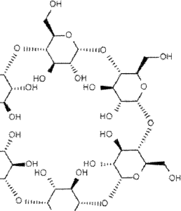
β-CYCLODEXTRIN
7 glucopyranose units; a medium cavity, approximately 6.0–6.5 Å.
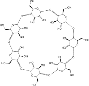
γ-CYCLODEXTRIN
8 glucopyranose units; the largest cavity, approximately 7.5–8.3 Å.
Cavity size affects which guests enter and how they are accommodated. In cited vitamin A tests, the larger γ-cyclodextrin cavity provided greater protection than β-cyclodextrin, making cavity geometry an important formulation variable.
After 48 days of UVA stress, a retinol/γ-CD complex retained 18.0% retinol, while the corresponding β-CD complex retained none (US 5,985,296, Example 3, Table 1).
Structure figures: US 8,410,079 B2 (2013), Figures 1–3. Cavity dimensions: Loftsson T, Brewster ME. J Pharm Sci. 1996;85(10):1017–1025.
PART II · THE SCIENCE OF CRYSTAL RETINAL
Molecular inclusion: a protective environment for retinal
HOST–GUEST ASSOCIATION: SCHEMATIC
Cyclodextrin host: hydrophilic exterior
Hydrophobic
cavity
Retinal guest
Noncovalent association: the guest is accommodated, not chemically modified.
DYNAMIC EQUILIBRIUM
Included retinal ⇌ Free retinal. As free retinal is taken up by skin during use, the equilibrium replenishes available active.
HOST–GUEST ASSOCIATION
Hydrophobic and van der Waals interactions drive lipophilic guests into the nonpolar cavity.
REDUCED ENVIRONMENTAL EXPOSURE
The included fraction has reduced exposure to bulk solvent, oxygen and light, key contributors to retinal degradation.
DISPERSION AND HANDLING
A lipophilic crystalline active becomes a water-dispersible solid ingredient for easier weighing, dispersion and processing.
EQUILIBRIUM-DRIVEN AVAILABILITY
Guest release follows the association equilibrium without shell rupture or a triggered chemical reaction.
DISTINCT FROM COVALENT DERIVATIVES
Inclusion is a physical association. The retinal–cyclodextrin hemiacetals discussed in the patent section are covalently bonded molecules of another class.
Loftsson T, Brewster ME. J Pharm Sci. 1996;85(10):1017–1025. Yap KL et al. Eur J Pharm Sci. 2005;25(1):49–56. Schematic for illustration; partial inclusion geometries of polyene guests have been reported.
PART II · THE SCIENCE OF CRYSTAL RETINAL
Stability through molecular design: interpreting stress tests
RETINOL RETENTION DURING OPEN STORAGE IN DAYLIGHT AT ROOM TEMPERATURE
Intact retinol measured by HPLC relative to day 0. US 5,985,296, Example 4, Table 2.
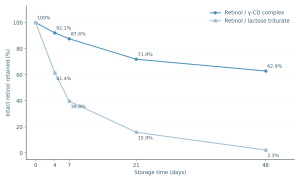
UVA AT 366 NM FOR 48 DAYS
18.0% retinol retained in the γ-CD complex
0% retained in the β-CD complex under the same conditions
US 5,985,296, Example 3, Table 1.
Protection depends on the molecular environment. In the patent comparisons, the γ-cyclodextrin complex retained most retinol after three weeks of open daylight storage, whereas the lactose triturate lost almost all of it.
Under UVA stress, γ-CD outperformed the smaller β-CD cavity, indicating the importance of inclusion geometry.
These data concern retinol, the immediate precursor of retinal, which shares related conjugated degradation chemistry.
US 5,985,296 A (Wacker-Chemie, 1999), Examples 3–4, Tables 1–2. Stability is complex-specific. These results characterize the tested materials and do not represent a particular commercial ingredient.
PART II · THE SCIENCE OF CRYSTAL RETINAL
Four decades of retinal and cyclodextrin patent science
1983
WATER-SOLUBLE RETINOID COMPLEXES
Retinal–polymer conjugates with cyclodextrin for water-compatible vitamin A systems.
US 4,371,673 (Pitha): an early approach to water-compatible retinoid delivery.
1999
γ-CD INCLUSION FOR VITAMIN A STABILITY
Quantified improvements in light and storage stability of retinol/γ-CD complexes.
US 5,985,296 (Wacker-Chemie): HPLC comparisons with β-CD and lactose controls.
2013
RETINAL–CD HEMIACETAL DERIVATIVES
Covalent retinal–cyclodextrin structures developed for skin-tone applications.
US 8,410,079 B2: chemically distinct from noncovalent inclusion complexes.
2020
MODERN RETINAL STABILIZATION
Cyclodextrin-encapsulated retinal combined with antioxidant systems.
US 10,864,148 B2: thermal and UV stress tests of encapsulated retinal gels.
WHAT THE PATENT HISTORY SHOWS
Successive approaches address retinoid instability through solubilization, inclusion, covalent derivatives and integrated formulations. Cyclodextrin inclusion is a route that protects the active without changing its molecular identity.
General retinal–cyclodextrin acetal structure
CN 103987391 B, Formula (Ib).
Exocyclic retinal–CD hemiacetal (VIII)
EP 2 755 664 B1 / HK 1200172 B, page 8.
US 4,371,673 A (1983) · US 5,985,296 A (1999) · US 8,410,079 B2 (2013) · US 10,864,148 B2 (2020).
PART II · THE SCIENCE OF CRYSTAL RETINAL
Comparing retinal formulation approaches
Three approaches to retinal formulation involve different practical considerations. This table compares formulation principles rather than ranking performance.
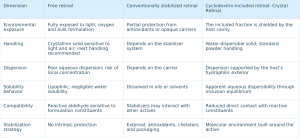
This is a qualitative comparison of formulation principles. Actual performance depends on the complex, manufacturing process and finished formulation. Validation parameters must be established during product development.
PART III · RETINAL BIOLOGY AND SKIN BENEFITS
From retinal to skin renewal: the signaling cascade
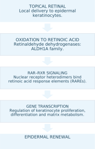
DOCUMENTED IN SKIN
RETINAL INCREASES CUTANEOUS RETINOIC ACID
Topical retinal increased retinoic acid content and retinoid activity in mouse skin, supporting enzymatic conversion within tissue (Didierjean et al., 1996; Sorg et al., 1999).
HUMAN EPIDERMAL RENEWAL MARKERS
In human skin, topical retinal induced epidermal hyperplasia and increased dermal elastin with good tolerability, consistent with retinoid-mediated renewal (Saurat et al., 1994).
RECEPTOR SPECIFICITY
In human keratinocytes, retinoid signaling at physiological, nanomolar ligand concentrations acts mainly through RAR–RXR heterodimers at βRARE elements (Xiao et al., 1995).
DOWNSTREAM VISIBLE ENDPOINTS
Texture, wrinkles, firmness, tone and barrier quality are mapped to human evidence in the following sections.
Didierjean L et al. J Invest Dermatol. 1996;107(5):714–719. Sorg O et al. Dermatology. 1999;199(Suppl 1):13–17. Saurat JH et al. J Invest Dermatol. 1994;103(6):770–774. Xiao JH et al. J Biol Chem. 1995;270(7):3001–3011.
PART III · RETINAL BIOLOGY AND SKIN BENEFITS
Skin texture: measurable renewal
Skin-surface refinement is an instrumentally measurable retinoid endpoint. Retinal supports orderly keratinocyte renewal, and changes can be assessed by profilometry and 3D imaging.
WHY THE SURFACE RESPONDS
Retinoic acid generated in skin modulates keratinocyte differentiation and epidermal turnover. Replacement of accumulated, disorganized corneocytes produces a more regular surface, measured as lower roughness in 3D imaging.
Retinal’s single-step conversion provides a rationale for renewal at cosmetic use concentrations.
+13.7%
Texture improvement with 0.1% retinal
+12.6%
Texture improvement with 0.05% retinal
Randomized double-blind trial: 40 women with photoaged skin, twice-daily use for 3 months; texture quantified by Antera 3D imaging.
BENCHMARK PROFILOMETRY STUDY
Creidi et al. J Am Acad Dermatol. 1998.
125 participants · 44 weeks · 0.05% retinal
DESIGN
Double-blind comparison of 0.05% retinal, 0.05% retinoic acid and vehicle in photoaged skin, with objective surface profilometry.
RESULTS
Both vitamin A groups reduced wrinkles and roughness versus vehicle, with significant improvement from week 18. Efficacy did not differ significantly between the active groups.
TOLERABILITY
Retinal was significantly better tolerated than retinoic acid at the same concentration.
For formulation development, these results support retinal as a cosmetic option with surface efficacy comparable to retinoic acid under the tested conditions. Inclusion provides a stabilization rationale that must be verified in the formulation.
Kwon HS et al. J Cosmet Dermatol. 2018;17(3):471–476. Creidi P et al. J Am Acad Dermatol. 1998;39(6):960–965. Studies used finished creams; results characterize the retinal active class.
PART III · RETINAL BIOLOGY AND SKIN BENEFITS
Wrinkles and fine lines: clinical evidence
TWELVE WEEKS, TWO CONCENTRATIONS, FOUR ENDPOINTS
Percentage change from baseline; randomized double-blind trial; 40 women; twice-daily use.
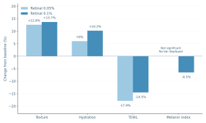
All displayed changes were significant versus baseline except the 0.05% group’s melanin endpoint, which was not significant and is therefore shown without a bar. Lower TEWL and melanin index indicate improvement.
LONG-TERM CONTROLLED EVIDENCE
Week 18: significant reduction versus vehicle
44-week follow-up; n = 125
Creidi P et al. J Am Acad Dermatol. 1998.
At the same 0.05% concentration, retinal provided wrinkle improvement comparable to retinoic acid by profilometry, with significantly less irritation.
The response persisted through the 44-week observation period, supporting a sustained rather than transient surface effect.
DOSE-SELECTION RATIONALE
In the reported results, 0.1% showed greater texture and pigment changes, while 0.05% showed the greater TEWL reduction. These endpoints inform different formulation concepts.
Kwon HS et al. J Cosmet Dermatol. 2018;17(3):471–476 (randomized double-blind trial, n = 40, 3 months; Antera 3D, corneometry, TEWL and pigment measurement). Creidi P et al. J Am Acad Dermatol. 1998;39(6):960–965 (n = 125, 44 weeks; profilometry).
PART III · RETINAL BIOLOGY AND SKIN BENEFITS
Dermal matrix: collagen and elastic-fiber support
Photoaging involves disrupted collagen and elastic fibers. Histology in skin explants and instrumental measurements in vivo provide evidence relevant to retinal and matrix-related care.
HUMAN SKIN EXPLANTS: UVA REPAIR MODEL
Fibers restored toward unexposed levels
Human skin explants were photoaged with UVA in organ culture and treated with 0.05% retinal cream for 2 weeks. In all examined specimens, collagen and elastic fibers returned to unexposed-skin levels, and UVA-suppressed collagen synthesis normalized.
Boisnic S et al. Dermatology. 1999;199(Suppl 1):43–48.
CLINICAL STUDY: ULTRASOUND AND RHEOLOGY
Instrumentally measured thickness and elasticity
Topical 0.05% retinal significantly increased epidermal thickness and skin elasticity in aging skin, quantified by high-frequency ultrasound and rheology.
Diridollou S et al. Dermatology. 1999;199(Suppl 1):37–41.
p < 0.01
Significance reported for increased epidermal thickness and elasticity with 0.05% retinal.
These findings provide a rationale for firmness and density concepts alongside surface smoothing.
SIGNALING BEHIND THE STRUCTURE
RAR-mediated transcription in epidermal keratinocytes is the main documented pathway. Explant data show matrix recovery consistent with renewed collagen synthesis after UV damage.
Active retention through stabilization is a formulation prerequisite that requires product-level confirmation.
Boisnic S et al. Dermatology. 1999;199(Suppl 1):43–48 (human skin explants, UVA + 0.05% retinal, 2 weeks). Diridollou S et al. Dermatology. 1999;199(Suppl 1):37–41 (0.05% retinal; ultrasound and rheology endpoints).
PART III · RETINAL BIOLOGY AND SKIN BENEFITS
Skin tone and pigmentation
MELANIN INDEX AT 12 WEEKS
−6.5%
A significant reduction in the 0.1% retinal group in the randomized double-blind photoaging trial.
Kwon HS et al. J Cosmet Dermatol. 2018.
The pigment endpoint was significant at 0.1% and not significant at 0.05%. This supports a concentration-specific rather than universal skin-tone claim.
HOW RETINAL RELATES TO SKIN TONE
PIGMENT CLEARANCE THROUGH TURNOVER
Accelerated epidermal renewal replaces melanin-containing keratinocytes and reduces surface pigment residence.
REPORTED DEPIGMENTING ACTIVITY
A dedicated study in pigmentary disorders reported measurable depigmenting activity of topical retinal, supporting even-tone formulation development.
Sorg O et al. Dermatology. 2013;227(3):231–237.
THE ROLE OF PHOTOSTABILITY
The source proposes light-stable inclusion as a way to retain activity in tone-care products intended for daytime use. This performance must be established for the specific complex and formulation.
POSITIONING
Combine tone-care evidence with texture and tolerability endpoints to support an even, refined skin appearance. Final product claims require validation.
Kwon HS et al. J Cosmet Dermatol. 2018;17(3):471–476 (n = 40, 3 months; significant melanin reduction only at 0.1%). Sorg O et al. Dermatology. 2013;227(3):231–237.
PART III · RETINAL BIOLOGY AND SKIN BENEFITS
Refined skin: pores and blemish-related mechanisms
The source reports direct antibacterial activity of retinal against Cutibacterium acnes, formerly Propionibacterium acnes, distinguishing it from the retinol and retinoic acid comparators discussed.
IN VITRO: MINIMUM INHIBITORY CONCENTRATION
4–8 mg/L
Retinal MIC against three P. acnes reference strains.
COMPARATOR
>128 mg/L
Retinoic acid MIC against the same strains.
This activity is attributed to retinal’s aldehyde group, a property of the molecule rather than a finished product.
Pechère M et al. Dermatology. 1999;199(Suppl 1):29–31.
IN VIVO: SINGLE-BLIND RANDOMIZED STUDY
The source slide records the median viable-count decrease per cm² as “≈ −10² log” after 2 weeks of daily 0.05% retinal. Vehicle alone was ineffective.
The numerical notation is ambiguous and is retained as written rather than converted to a different unit.
COMPLEMENTARY EVIDENCE
COMEDOLYTIC ACTIVITY
Topical retinal reduced comedones in the rhino mouse model, providing a complementary mechanism for pore-focused concepts.
Fort-Lacoste L et al. Dermatology. 1999;199(Suppl 1):33–35.
Use as mechanistic support for clarifying and blemish-prone-skin positioning, rather than a standalone acne-treatment claim.
Pechère M et al. Dermatology. 1999;199(Suppl 1):29–31. Fort-Lacoste L et al. Dermatology. 1999;199(Suppl 1):33–35. In vitro and animal findings provide mechanistic support, not clinical acne efficacy.
PART III · RETINAL BIOLOGY AND SKIN BENEFITS
Barrier function, hydration and tolerability
BARRIER FUNCTION: KWON 2018 RANDOMIZED TRIAL
−14.5%
TEWL in the 0.1% group
−17.9%
TEWL in the 0.05% group
+10.2%
Skin hydration in the 0.1% group
+6.0%
Skin hydration in the 0.05% group
After 12 weeks of twice-daily retinal use, lower TEWL and greater stratum corneum hydration indicated improvements versus baseline.
TOLERABILITY UNDER CHALLENGE
MAXIMIZED AND LONG-TERM EXPOSURE
In a tolerability study with 355 volunteers under maximized and long-term conditions, retinoic acid caused significantly more erythema and scaling, while retinal was closer to vehicle.
Fluhr JW et al. Dermatology. 1999;199(Suppl 1):57–60.
NO PHOTOTOXICITY SIGNAL
A dedicated human tolerability review involving 357 subjects reported a favorable safety profile for topical retinal, including no phototoxic reactions.
Sachsenberg-Studer EM. Dermatology. 1999;199(Suppl 1):61–63.
MINIMAL SYSTEMIC EXPOSURE
Plasma retinoid levels were essentially unchanged after topical use in human skin, supporting local-action positioning.
Sass JO et al. Skin Pharmacol. 1996;9(5):322–326.
FORMULATION VALUE
Tolerability is important for retinoid adherence. The cited barrier and tolerability findings inform the development of everyday retinal products.
Kwon HS et al. J Cosmet Dermatol. 2018;17(3):471–476. Fluhr JW et al. Dermatology. 1999;199(Suppl 1):57–60. Sachsenberg-Studer EM. Dermatology. 1999;199(Suppl 1):61–63. Sass JO et al. Skin Pharmacol. 1996;9(5):322–326.
PART IV · SCALP SCIENCE AND HAIRCARE INNOVATION
The scalp is skin and responds to retinoid signaling
PuriActives® Crystal Retinal: retinal held within a cyclodextrin inclusion complex.
Scalp care draws on skincare concepts such as renewal, barrier support and the microbiome. The scalp is a specialized, follicle-rich skin surface with relevant retinoid biology.
A RETINOID-RESPONSIVE SURFACE
Scalp epidermis shares retinoid receptor mechanisms with facial skin. The retinal-to-retinoic-acid pathway can operate in keratinocytes across body sites.
A MICROBIAL DIMENSION
The follicle-rich scalp carries dense microbial communities, including C. acnes. Retinal’s reported activity against this organism provides a follicular-environment rationale.
FORMULATION CHALLENGES
Scalp products are often water-rich, surfactant-containing and transparent, creating stability challenges for unprotected retinal.
Retinoid skin biology: Saurat JH et al. J Invest Dermatol. 1994;103(6):770–774. Antibacterial activity: Pechère M et al. Dermatology. 1999;199(Suppl 1):29–31.
PART IV · SCALP SCIENCE AND HAIRCARE INNOVATION
Follicular biology: interpreting receptor evidence
The follicle is a retinoid-sensitive mini-organ. Receptor genetics defines the biological rationale and the limits of scalp-care positioning.
GENETIC EVIDENCE: RXRα AND THE HAIR CYCLE
Keratinocyte RXRα is required for a normal hair cycle.
Mice with targeted RXRα loss in epidermal and follicular keratinocytes developed progressive alopecia and failed to initiate anagen, establishing a requirement for this receptor signaling in the hair cycle.
Li M et al. Development. 2001;128(5):675–688.
REVIEW EVIDENCE: THE PILOSEBACEOUS UNIT
Retinoids act on follicular glands and epithelium.
An endocrine review of pilosebaceous biology discusses retinoid effects on sebocyte differentiation and follicular keratinization, processes relevant to scalp condition, oiliness and scaling.
Deplewski D, Rosenfield RL. Endocr Rev. 2000;21(4):363–392.
POSITIONING WITHIN THE EVIDENCE
Retinoid effects are dose-dependent, and excessive systemic exposure is associated with hair loss.
Crystal Retinal’s scalp positioning therefore concerns a balanced, comfortable scalp and follicular environment.
No hair-growth, density-increase or hair-loss-prevention claim is made or implied.
TAKEAWAY
Follicular retinoid sensitivity is established biology; cosmetic development opportunities concern scalp condition.
Li M et al. Development. 2001;128(5):675–688. Deplewski D, Rosenfield RL. Endocr Rev. 2000;21(4):363–392. Mechanistic evidence, not clinical validation of Crystal Retinal.
PART IV · SCALP SCIENCE AND HAIRCARE INNOVATION
Scalp-care opportunities from the formulation bench
Premium haircare increasingly uses skincare actives. Retinal development requires stability in realistic scalp formats: water-rich systems with surfactants and potentially prolonged residence.
THE CHALLENGE
Free retinal is difficult to stabilize in scalp formats.
Water, oxygen, light and surfactant environments can promote loss of unprotected retinal during product development and storage.
THE INCLUSION APPROACH
A protected guest available at the interface.
The inclusion complex reduces exposure of retinal’s conjugated system in the bulk formulation and allows guest exchange at the skin interface through water activity and lipid affinity.
A BRAND DEVELOPMENT CONCEPT
Skincare renewal adapted for the scalp.
Scalp comfort, balance, gentle renewal and follicular-environment care are anchored in published skin biology rather than hair-growth promises.
Stability engineering creates an opportunity to explore retinal in demanding scalp formats.
PART V · FORMULATION AND APPLICATIONS
Formulating Crystal Retinal: working principles
Inclusion chemistry provides a stabilization rationale, but processing remains important. Confirm each of these eight principles in the customer’s own stability program.
01 ADD DURING COOL-DOWN
Introduce the complex below typical hot-process temperatures to protect host–guest equilibrium.
02 CONTROL LIGHT FROM THE START
Use amber or opaque handling through production and filling.
03 BUILD ANTIOXIDANT SUPPORT
Chelators and antioxidant systems remain useful for retinoids, whether included or free.
04 SET pH USING MEASUREMENTS
Mildly acidic conditions are generally favorable; confirm the suitable window in the final base.
05 SCREEN COMPETING GUESTS
Hydrophobic fragrance constituents and oils may compete for the cyclodextrin cavity. Check displacement risk in compatibility tests.
06
AVOID STRONG OXIDATIVE CHALLENGES
Peroxides and aggressive preservative boosters can affect the polyene chain. Screen the full combination.
07
SPECIFY PROTECTIVE PACKAGING
Airless, low-permeability, light-protective packaging supports shelf-life retention.
08
VALIDATE IN THE REAL FORMULATION
Run accelerated and real-time stability tests on the finished product. Complex data guide development but do not replace product-level evidence.
Principles are drawn from retinoid and cyclodextrin literature. PuriPharm technical support can advise on specific bases.
Loftsson T, Brewster ME. J Pharm Sci. 1996;85(10):1017–1025. Yap KL et al. Eur J Pharm Sci. 2005;25(1):49–56. These principles are not validated specifications for a particular formulation.
PART V · FORMULATION AND APPLICATIONS
Skincare concepts informed by the evidence
NIGHT RENEWAL SERUM
Texture and wrinkle endpoints from randomized studies provide claim-development anchors. Inclusion is intended to support active retention during shelf life.
Evidence anchors: Kwon 2018; Creidi 1998.
GENTLE EVERYDAY RETINAL MOISTURIZER
Barrier findings and vehicle-like tolerability support routine-use development.
Evidence anchors: Fluhr 1999; Kwon 2018, TEWL and hydration.
FIRMING AND DENSITY CREAM
Ultrasound and explant evidence concerning thickness, elasticity and matrix recovery informs firmness concepts.
Evidence anchors: Diridollou 1999; Boisnic 1999.
EVEN-TONE SERUM
Significant melanin-index reduction at 0.1% and reported depigmenting activity support a concentration-specific concept.
Evidence anchors: Kwon 2018; Sorg 2013.
CLARIFYING CARE FOR BLEMISH-PRONE SKIN
C. acnes activity and comedolytic findings provide mechanistic support for pore-focused formulations.
Evidence anchors: Pechère 1999; Fort-Lacoste 1999.
EYE-AREA RENEWAL CARE
The source proposes retinal’s efficacy and tolerability profile as a development rationale for thin, sensitive skin around the eyes.
Evidence anchor: the cited tolerability literature.
Concepts only. Validate all claims in the final marketed product. Evidence anchors refer to the skin-benefit studies reviewed above.
PART V · FORMULATION AND APPLICATIONS
Scalp-care concepts
Four dosage-form concepts explore stabilized retinal for scalp care while retaining the limits of the evidence.
LEAVE-ON SCALP SERUM
A night-care concept for surface renewal and comfort, informed by retinal epidermal biology and tolerability literature.
BALANCING TONIC FOR OILY, CONGESTION-PRONE SCALPS
Reported C. acnes activity and follicular keratinization mechanisms support a clean, balanced follicular-environment concept.
INTENSIVE PRE-WASH CARE
A weekly concentrated step in an aqueous base, with active retention as a formulation-development objective.
CONDITIONER AND HAIR-MASK ADDITION
Explore rinse-off systems. Guest release and rinse compatibility require joint engineering by the brand and PuriPharm.
Concepts only. Use scalp-condition wording. The cited evidence does not support hair-growth, density-increase or hair-loss-prevention claims.
PART V · FORMULATION AND APPLICATIONS
Patent evidence: what the documents show
CN 103987391 B: a granted Chinese patent showing retinal–cyclodextrin acetal conjugate structures.
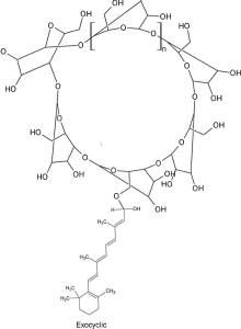
CN 103987391 B patent family: exocyclic retinal–cyclodextrin hemiacetal structure (VIII).
CN 103987391 B patent family: endocyclic structure (VII).
INTERPRETING THE RECORD
Patent documents cover both noncovalent inclusion complexes and covalent conjugates. PuriActives® Crystal Retinal is positioned as a molecular inclusion complex based on physical equilibrium. Related patent test data are discussed in the stability and patent-history sections.
Figures reproduced from granted patent documents for technical discussion: CN 103987391 B; the source also cites the US 10,864,148 B2 family. Patent structures and comparisons are not product specifications for Crystal Retinal.
PART VI · COMMERCIAL POSITIONING
One ingredient, four value pillars
MOLECULAR STABILIZATION
Cyclodextrin inclusion reduces environmental exposure of retinal’s conjugated system, informed by host–guest chemistry and retinoid-stabilization patents.
EFFICACY INFORMED BY RETINOIC ACID COMPARISONS
One metabolic step from the active form, with randomized cosmetic-concentration studies on texture, wrinkles, barrier function, hydration and pigmentation.
FORMULATION FLEXIBILITY
Inclusion enables exploration of creams, aqueous serums and scalp formats, with guest release at the skin interface as the development rationale.
A DOCUMENTED SCIENTIFIC STORY
Peer-reviewed clinical literature, patent tests and receptor biology provide a framework for claim development and review.
PRODUCT & PARTNERSHIP
Retinal science. The possibilities of molecular inclusion.
PuriActives® Crystal Retinal Cyclodextrin-Included Retinal combines a direct cosmetic retinoid precursor with molecular stabilization for skin and scalp formulation development.
PuriPharm Co., Ltd.
PuriActives® Crystal Retinal · INCI: RETINAL
Samples, technical documentation and development support available on request.
www.puriactives.com
This report is intended for professionals and summarizes published research and granted patent documents. Data characterize the retinal active class and the cited test materials. Finished-product claims require product-level validation.
NanoActive Oleu™
NanoActive Oleu™

PuriPharm Co., Ltd. · Technical Report
Liposomal Oleuropein
Mediterranean polyphenol defense
Enhanced by delivery science
INCI: OLEA EUROPAEA (OLIVE) LEAF EXTRACT
Powered by PURISOME® FlexVes™ Liposome Technology
01 · INGREDIENT OVERVIEW
NanoActive Oleu™ at a glance
PRODUCT NAME:NanoActive Oleu™ Liposomal Oleuropein
INCI NAME:Olea Europaea (Olive) Leaf Extract
BOTANICAL SOURCE:Leaves of Olea europaea L.
KEY PHYTOCHEMICAL:Oleuropein, a secoiridoid polyphenol
DELIVERY PLATFORM:PURISOME® FlexVes™ Liposome Technology
PHYSICAL FORM:Liposomal dispersion for aqueous and emulsified systems
APPLICATION AREAS:Skincare · Oral care · Hair and scalp care
PRODUCT POSITIONING:An oleuropein-rich olive leaf extract encapsulated in flexible liposomal vesicles. NanoActive Oleu™ brings Mediterranean polyphenol science to formulation-ready skin, oral and scalp care concepts, informed by human, in vivo and mechanistic research.
Young olive leaves (Olea europaea L.), the botanical source of the actives.
02 · BOTANICAL ORIGIN
Olive leaves: a concentrated source of polyphenols
Olive branch with young leaves and fruit. Leaves are the tree’s richest source of phenolic compounds.
60–90 mg oleuropein per g of dry leaves
The olive tree concentrates its defensive chemistry in its leaves. Oleuropein, the predominant secoiridoid polyphenol, can reach 60–90 mg/g of dry leaves, alongside hydroxytyrosol, verbascoside and flavonoid glycosides.
This phytochemical system evolved under sunlight, microbial and oxidative stress, providing a botanical rationale relevant to daily challenges faced by skin, oral mucosa and scalp.
Nediani C, et al. Antioxidants. 2019;8(12):578. doi:10.3390/antiox8120578
Omar SH. Sci Pharm. 2010;78(2):133–154.
03 · KEY ACTIVE MOLECULE
Oleuropein: secoiridoid chemistry for redox activity
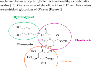
Oleuropein (molecular weight 540.5): hydroxytyrosol + elenolic acid + glucose. Nediani C, et al. Antioxidants. 2019;8:578.
CATECHOL CORE
The catechol hydroxyls of the hydroxytyrosol moiety donate hydrogen atoms to neutralize free radicals.
ACTIVE PRODUCTS OF HYDROLYSIS
Ester hydrolysis releases hydroxytyrosol and elenolic acid, extending the molecule’s functional profile.
NATURAL HYDROPHILICITY
A water-soluble glycoside with biological activity, but limited passage through lipid-rich barriers without delivery support.
This chemistry provides the rationale for complementary delivery engineering.
04 · DELIVERY CHALLENGES
Active polyphenols face practical delivery challenges
CHEMICAL INSTABILITY
Oleuropein’s ester bonds and catechol groups are susceptible to hydrolysis and oxidation, potentially reducing activity during shelf life.
BARRIER MISMATCH
Hydrophilic glycosides partition poorly into lipid-rich stratum corneum and mucosal surfaces, limiting local bioavailability.
SHORT RESIDENCE TIME
Unencapsulated polyphenols can be diluted, metabolized or washed away quickly, particularly in the oral environment.
FORMULATION CONSTRAINTS
Oxidation-sensitive polyphenols constrain pH, processing, packaging and preservative-system design.
DELIVERY APPROACH
Delivery science aims to turn a fragile molecule into a robust, formulation-ready ingredient.
PURISOME® FlexVes™ Liposome Technology
The proprietary liposomal platform behind the NanoActive™ ingredient range is introduced below.
Stability reference: Perugini P, et al. Int J Cosmet Sci. 2008;30(2):113–120.
05 · DELIVERY PLATFORM
PURISOME® FlexVes™ Liposome Technology
Flexible phospholipid vesicles: PuriPharm’s delivery architecture.
PuriPharm’s proprietary platform encapsulates oxidation-sensitive polyphenols in flexible liposomal vesicles, with four proposed formulation benefits.
01 · PROTECTION
Protect actives vulnerable to oxidation and degradation to support shelf life.
02 · PENETRATION
Promote stratum corneum penetration and improve bioavailability.
03 · RELEASE
Provide sustained, controlled release to reduce irritation risk.
04 · COMPATIBILITY
Improve formulation compatibility and skin feel.
PuriPharm Co., Ltd. has more than ten years of liposome R&D experience.
06 · INTEGRATED VALUE
One ingredient, a complete value framework
01
OLIVE LEAVES
BOTANICAL SOURCE
Olea europaea L. leaves, the tree’s most polyphenol-rich plant part.
02
PHYTOCHEMISTRY
ACTIVE FOUNDATION
An oleuropein-rich polyphenol system with published evidence across multiple pathways.
03
PURISOME®
DELIVERY ENGINEERING
FlexVes™ liposomal protection, penetration and controlled release.
04
FORMULATION FIT
FLEXIBLE USE
Dispersion for incorporation into emulsions, gels, serums and oral-care bases.
05
SKIN · ORAL · SCALP
APPLICATION AREAS
Three care areas, each with its own relevant evidence base.
Botanical chemistry informs delivery engineering, delivery supports formulation design, and formulations translate the concept into three care areas.
07 · MECHANISM OVERVIEW
Five pillars of cellular defense
WHY FIVE PILLARS?
UV exposure, pollution, microorganisms and mechanical stress can occur together and converge on shared cellular mechanisms.
Oleuropein research addresses this network from radical scavenging to cellular quality control.
Mechanisms and supporting evidence are reviewed below.
REDOX DEFENSE
Direct radical scavenging and support for endogenous antioxidant systems.
INFLAMMATORY MODULATION
Modulation of NF-κB-related signaling and key inflammatory mediators.
CELLULAR QUALITY CONTROL
Autophagy and proteostasis pathways involving AMPK, mTORC1, SIRT1 and TFEB.
TISSUE REPAIR
Support for re-epithelialization, collagen deposition and VEGF signaling.
MICROBIAL BALANCE
Published inhibitory activity against biofilm-forming, cariogenic oral bacteria.
Mechanistic framework: Nediani C, et al. Antioxidants. 2019;8(12):578.
07 · MECHANISM: REDOX DEFENSE
Quantifying radical-scavenging activity
IN VITRO EVIDENCE
Oleuropein scavenges DPPH radicals in a concentration-dependent manner, with a curve approaching that of the reference antioxidant ascorbic acid. Complementary ABTS assays also support anti-radical activity.
CHEMICAL RATIONALE
· Catechol hydroxyl groups donate hydrogen atoms to neutralize radicals.
· The resulting ortho-quinone structure is resonance-stabilized.
· Hydrolysis releases hydroxytyrosol, contributing further activity.
In vitro radical-scavenging activity provides a rationale for using PURISOME® delivery to improve contact with sites of UV- and pollution-related oxidative stress.
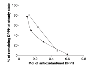
DPPH radical-scavenging activity: oleuropein (●) versus ascorbic acid (○, reference). Perugini P, et al. Int J Cosmet Sci. 2008;30(2):113–120.
Dose-dependent radical scavenging is the first mechanistic step, upstream of inflammation, barrier damage and visible aging.
07 · MECHANISM: INFLAMMATORY MODULATION
Interrupting the oxidative stress–inflammation cycle
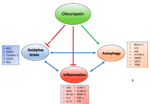
Oxidative stress–autophagy–inflammation network. The indicated molecular markers are discussed in the oleuropein literature. Adapted from Nediani C, et al. Antioxidants. 2019;8(12):578.
NF-κB SIGNALING
Modulated upstream of cytokine-cascade amplification.
COX-2 AND iNOS
These inflammatory enzymes are downregulated in experimental models.
IL-6 · TNF-α · IL-1β
Key inflammatory mediators are reduced in cellular and tissue studies.
ICAM-1 · VCAM-1
Reduced adhesion-molecule expression is associated with less immune-cell recruitment and inflammation.
Oleuropein acts at the oxidative entry point of the cycle, providing an upstream intervention rationale.
07 · MECHANISM: CELLULAR QUALITY CONTROL
Autophagy as a quality-control mechanism
Autophagy is the cellular recycling system that identifies and clears damaged proteins and organelles. Oleuropein has been reported as an autophagy inducer in several experimental models.
01
STRESS SENSING
AMPK activation increases, while the mTORC1 brake on autophagy is reduced.
AMPK ↑ · mTORC1 ↓
02
AUTOPHAGY INITIATION
Beclin-1 participates; LC3-I converts to LC3-II as autophagosomes form.
Beclin-1 · LC3-II ↑
03
SUBSTRATE CLEARANCE
p62 directs damaged proteins and organelles toward lysosomal degradation.
p62 turnover ↑
04
CELLULAR RENEWAL
SIRT1 and TFEB support lysosomal biogenesis and cellular renewal.
SIRT1 · TFEB ↑
This cellular clearance mechanism provides a distinctive basis for botanical active development.
Autophagy markers: Nediani C, et al. Antioxidants. 2019;8(12):578. doi:10.3390/antiox8120578
07 · MECHANISM: PROTEOSTASIS
Proteostasis support for lasting skin quality
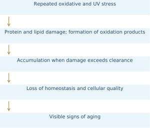
THE PROTEOSTASIS PERSPECTIVE
The proteostasis network, including synthesis, folding, repair and degradation, maintains functional proteins. Under chronic environmental stress, damaged material can accumulate faster than cells can clear it.
Oleuropein is positioned on both sides of this balance: antioxidant defense slows damage formation, while autophagy induction supports clearance.
Formulation implication: ingredients that support cellular quality control provide a rationale for long-term skin resilience as well as repair.
Proteostasis and autophagy framework: Nediani C, et al. Antioxidants. 2019;8(12):578.
08 · SKINCARE: HUMAN EVIDENCE
Human evidence under UVB stress
STUDY DESIGN
MODEL
UVB-induced erythema in human volunteers, a controlled photobiology challenge model.
TEST FORMULATIONS
Topical oleuropein emulsions and emulgels applied after UVB exposure.
REFERENCE
Vitamin E, a topical antioxidant benchmark.
OBJECTIVE ENDPOINTS
· Erythema index: reflectance colorimetry
· TEWL: transepidermal water loss
· Skin blood flow: laser Doppler flowmetry
WHY THE DESIGN MATTERS
The controlled UVB challenge uses objective biophysical measurements in human volunteers rather than self-assessment.
It evaluates whether the test formulations can help limit redness and preserve barrier function after a defined oxidative challenge.
Perugini P, et al. Int J Cosmet Sci. 2008;30(2):113–120. doi:10.1111/j.1468-2494.2007.00416.x
08 · SKINCARE: HUMAN EVIDENCE
Approximately 35% lower TEWL after UVB exposure
–35%
TEWL: measurable preservation of barrier function
–22%
Erythema index: reduced visible redness
–30%
Skin blood flow: reduced hyperemic response
Approximate mean reductions versus the UVB-exposed control, for oleuropein emulsions and emulgels. Perugini P, et al. Int J Cosmet Sci. 2008;30(2):113–120.
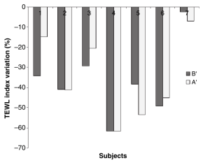
Transepidermal water loss (TEWL)
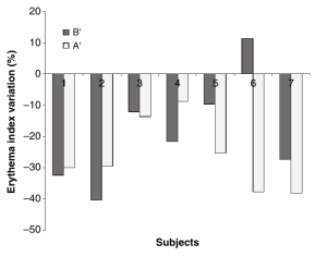
Erythema index (colorimetry)
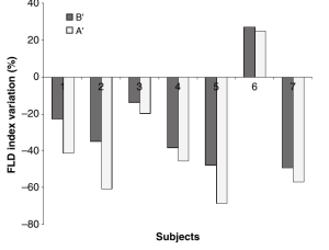
Blood flow (laser Doppler)
Three instrumental endpoints support reduced redness, barrier disruption and vascular response under the tested conditions.
08 · SKINCARE: HUMAN EVIDENCE
From acute photostress to photoaging care
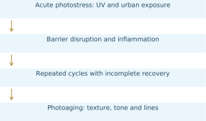
The controlled UVB study provides a rationale for daily photoaging care. Longer-term human data on an olive leaf extract cream extend the evidence beyond an acute challenge.
LONGER-TERM HUMAN DATA: OLIVE LEAF EXTRACT CREAM
36 female volunteers; twice daily for 2 months:
· TEWL decreased significantly (p = 0.007), with the effect maintained 1 month after discontinuation.
· Skin hydration increased (p = 0.004).
· Wrinkle parameters improved (p < 0.001).
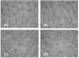
Visioscan® skin surface images: baseline (A), 1 month (B), 2 months (C), and 1 month after discontinuation (D).
Wanitphakdeedecha R, et al. J Cosmet Dermatol. 2020. doi:10.1111/jocd.13457
08 · SKINCARE: PRECLINICAL EVIDENCE
Tissue repair and matrix renewal
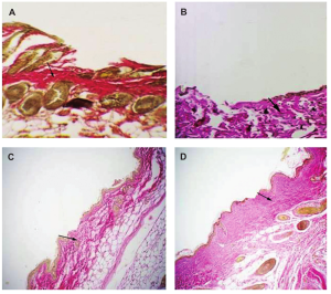
Van Gieson staining of collagen deposition in healing skin: oleuropein-treated versus control groups. Mehraein F, et al. Cell J. 2014;16(1):25–30.
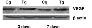
VEGF protein expression by Western blot: oleuropein group (Tg) versus control group (Cg).
Increased VEGF supports microvascular supply to repairing tissue and provides a mechanistic link with matrix renewal.
RESULTS IN AN AGED-SKIN MODEL
Wound closure at day 3: 29.0% versus 19.2% in controls.
At day 7: 55.3% versus 45.3% in controls.
Re-epithelialization was complete by day 7 in treated skin.
Collagen deposition increased; VEGF increased (p < 0.05).
Patent support: EP2570124A1 covers oleuropein compositions for repair of aged and fragile skin.
Intradermal model in aged mice: preclinical evidence, not a human efficacy claim.
08 · SKINCARE: HUMAN AND IN VITRO EVIDENCE
Even skin tone: observations in melanin biology
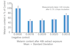
Melanin content in primary human melanocytes (in vitro).
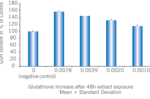
Intracellular glutathione (in vitro).
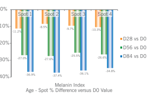
Age-spot melanin index versus baseline (D0) in a 12-week human study.
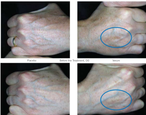
Placebo versus test product at week 12.
IN VITRO: HUMAN MELANOCYTES
Melanin content decreased by up to approximately 65%, while intracellular glutathione increased by 55%, supporting the cellular redox-buffering system.
HUMAN STUDY: AGE SPOTS
In a 12-week double-blind, placebo-controlled study of 12 women aged 46–72 years, the age-spot melanin index decreased by 34.8% at week 12 versus baseline, with visible lightening.
The source connects skin-tone care with the redox rationale used for barrier defense.
Standardized olive polyphenol extract. Lueder M. Cosmet Sci Technol. 2011.
09 · ORAL CARE
The mouth: a redox–microbial ecosystem
Biofilms reform within hours, saliva continuously dilutes actives, and soft tissues face daily mechanical, chemical and microbial stress. Oral care therefore requires both suitable chemistry and effective delivery.
BIOFILM AND PLAQUE
Bacterial biofilms drive plaque accumulation and acid challenges. Daily biofilm control provides an application opportunity for polyphenols.
GINGIVAL ENVIRONMENT
The gingival margin lies at the intersection of microbial and oxidative stress. Redox support and inflammatory modulation provide relevant development pathways.
SOFT-TISSUE REPAIR
Mucosal tissues face daily irritation and discomfort after care procedures. Repair support is a relevant consumer-care concept.
Three application opportunities share the same polyphenol rationale: antioxidant activity, inflammatory modulation and microbial inhibition.
09 · ORAL CARE: IN VITRO EVIDENCE
Published activity against key oral bacteria
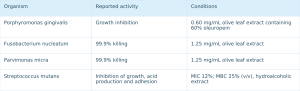
In vitro data support a biofilm-management concept and do not constitute therapeutic claims.
Karygianni L, et al. Biomed Res Int. 2014;2014:839019. doi:10.1155/2014/839019.
S. mutans: Golestannejad Z, et al. Dent Res J (Isfahan). 2020;17(3):179–185. doi:10.4103/1735-3327.284730 (acid production and adhesion: p < 0.001).
09 · ORAL CARE: HUMAN EVIDENCE
Biofilm management beyond disinfection
PLAQUE REGROWTH: CROSSOVER CONTROLLED STUDY, N = 17
36% lower plaque index versus water rinsing
After rinsing with a virgin olive oil preparation containing oleuropein, the plaque index was 1.48 ± 0.36 versus 2.30 ± 0.39 with water (p < 0.001), measured in a plaque-regrowth crossover design.
Volunteers suspended oral hygiene and underwent objective plaque-regrowth scoring, providing a direct measure of biofilm management under the study conditions.
Math MV, et al. Int J Dent Health Sci. 2017;4(4).
THE POLYPHENOL RATIONALE FOR BIOFILM CARE
INTERFERING WITH ADHESION
Olive leaf polyphenols reduce S. mutans surface adhesion (p < 0.001).
MODULATING ACID PRODUCTION
Reduced acid production by cariogenic bacteria supports a daily-care rationale relevant to enamel.
REDOX SUPPORT AT THE GINGIVAL MARGIN
Antioxidant activity at the biofilm–gingiva interface provides a rationale for comfort-oriented care.
Golestannejad Z, et al. Dent Res J (Isfahan). 2020;17(3):179–185.
09 · ORAL CARE: CLINICAL AND PRECLINICAL EVIDENCE
Oral soft-tissue defense: cytokines and comfort
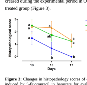
Mucosal histopathology scores over time: oleuropein versus controls in a mucositis model.
Tissue MDA, a marker of oxidative damage: oleuropein versus controls (reported p = 0.000).
CLINICAL: RANDOMIZED CROSSOVER TRIAL, 25 PATIENTS
Olive leaf extract mouthwash reduced salivary IL-1β and TNF-α and was associated with lower mucositis severity scores in chemotherapy patients.
PRECLINICAL: MUCOSITIS MODEL
Oleuropein ointment reduced histopathology scores and tissue MDA (reported p = 0.000), indicating less tissue damage and oxidative stress in the experimental mucosal challenge.
Positioning: supportive oral care and comfort. Use adjunctive wording rather than therapeutic claims.
Ahmed KM. Saudi Dent J. 2013;25(4):141–147. doi:10.1016/j.sdentj.2013.09.001.
Showraki N, et al. J Dent (Shiraz). 2016;17(4):334–342.
09 · ORAL CARE: DELIVERY SCIENCE
Why delivery matters in oral care
FOUR ORAL-ENVIRONMENT CHALLENGES
Continuous dilution: saliva washes actives away.
Short contact time: rinsing lasts minutes rather than hours.
Oxidative exposure: polyphenols are vulnerable to degradation in aqueous systems.
Dosage-form constraints: clear rinses, gels and pastes have different formulation requirements.
THE PURISOME® APPROACH
· Vesicles protect polyphenols within the formulation.
· Controlled release aims to extend availability after use.
· The dispersion format suits rinses, gels and pastes.
SUPPORTING LITERATURE
Liposomal encapsulation of olive leaf polyphenols preserved, and under some conditions enhanced, antibacterial activity against S. aureus, including MRSA (MIC 140–240 µg/mL). Vesicles remained stable for up to 90 days at 4 °C.
Prevete G, et al. ACS Appl Mater Interfaces. 2024;16(50):68850–68863. doi:10.1021/acsami.4c13302
10 · HAIR AND SCALP CARE
The scalp: a stressed follicular environment
The scalp is skin with approximately 100,000 follicles, abundant sebum and direct UV exposure, creating a concentrated set of environmental challenges.
PHOTOSTRESS
UV exposure is particularly relevant at partings and areas of thinning hair.
SEBUM OXIDATION
Lipid peroxidation at follicular openings increases the oxidative load.
MICROBIAL PRESSURE
A dense, sebum-supported microbial community contributes to ongoing surface challenges.
FOLLICULAR CYCLE
Transition into anagen is sensitive to oxidative and inflammatory signaling.
Scalp care can draw on the same defensive mechanisms relevant to skin care.
10 · HAIR AND SCALP CARE
Oleuropein as a rationale for scalp resilience
REDOX DEFENSE
Radical scavenging at the scalp surface, where sebum oxidation and UV exposure converge.
Supporting evidence: in vitro and human skin data.
INFLAMMATORY MODULATION
A rationale for perifollicular comfort through modulation of inflammatory mediators.
Supporting evidence: mechanistic studies and human oral-care data.
MICROBIAL BALANCE
Published antibacterial activity against biofilm-forming organisms.
Supporting evidence: in vitro MIC data.
QUALITY CONTROL
Autophagy support as a concept for rapidly dividing cells in the follicular environment.
Supporting evidence: mechanistic literature.
FOLLICULAR SIGNALING
Wnt/β-catenin activation in dermal papilla cells, detailed below.
Supporting evidence: preclinical data.
Each scalp-care pillar is presented with its supporting evidence type.
10 · HAIR AND SCALP: PRECLINICAL EVIDENCE
Follicular biology: Wnt/β-catenin activation
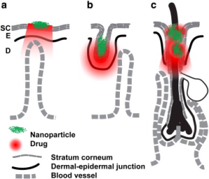
Nanovesicle enrichment through the follicular route: a PURISOME® delivery concept.
HUMAN DERMAL PAPILLA CELLS: IN VITRO
Oleuropein promoted dermal papilla cell proliferation, increased LEF1 and Cyclin D1, and drove nuclear β-catenin accumulation, consistent with Wnt pathway activation.
TELOGEN MOUSE MODEL: TOPICAL APPLICATION FOR 28 DAYS
Anagen entry accelerated, with larger and more numerous follicles. Wnt10b, FzdR1, LRP5, IGF-1, KGF, HGF and VEGF increased in skin tissue.
Evidence level: preclinical. These findings support scalp-care positioning and further development, not a human hair-growth claim.
Tong T, Kim N, Park T. PLoS ONE. 2015;10(6):e0129578. doi:10.1371/journal.pone.0129578
11 · APPLICATIONS
Three application areas, one technology platform
SKINCARE
DEFENSE AND REPAIR
· Human UVB study: TEWL –35%; erythema –22%.
· Photoaging care: TEWL p = 0.007; wrinkles p < 0.001.
· Even tone: age-spot melanin index –34.8%.
· Tissue repair: preclinical wound models.
ORAL CARE
BIOFILM AND SOFT-TISSUE BALANCE
· Antibacterial activity: published MIC/MBC values.
· Human crossover study: plaque regrowth –36%.
· Clinical rinse study: reduced salivary IL-1β and TNF-α.
· Mucosal comfort: preclinical mucositis model.
HAIR AND SCALP
RESILIENCE AND FOLLICULAR SUPPORT
· Wnt/β-catenin activation: preclinical.
· Follicular delivery: PURISOME® concept.
· Redox and microbial rationale: evidence from skin and oral studies.
One ingredient concept connects three care areas through a shared scientific rationale.
11 · APPLICATIONS
From ingredient to formulation: product concepts
SKINCARE
Daily defense serum
After-sun repair emulgel
Even-tone concentrate
Barrier-support lotion
Informed by human UVB, photoaging and skin-tone studies.
ORAL CARE
Daily defense mouthwash
Gingival-care gel
Biofilm-management toothpaste
Soothing rinse after care procedures
Informed by antibacterial, plaque and cytokine evidence.
HAIR AND SCALP
Pre-wash scalp concentrate
Leave-on scalp serum
Clarifying antioxidant shampoo
Informed by preclinical follicular data and the scalp-stress rationale.
These are concepts for joint development. Final product claims require testing of the finished formulation.
11 · APPLICATIONS
NanoActive Oleu™ formulation guidance
HANDLING PRINCIPLES
Supplied as an aqueous liposomal dispersion, with no reconstitution required.
Add during cool-down. Avoid prolonged high shear to protect vesicle integrity.
Suitable formulation areas include emulsions, serums, gels and oral-care systems.
Use mild temperatures and pH conditions; validate the preservative system in the complete formulation.
Detailed specifications, recommended use levels and starter formulations are available from PuriPharm technical services.
WORKING WITH PURIPHARM
Ingredient supply with joint development support.
PuriPharm provides application expertise, formulation guidance and supporting evidence documentation, from the first prototype to market-registration materials.
Contact PuriPharm Co., Ltd. for technical services and joint development.
NANOACTIVE OLEU™ LIPOSOMAL OLEUROPEIN
Protect · Balance · Renew
PHYTOCHEMISTRY: oleuropein-rich Mediterranean polyphenols
DELIVERY: PURISOME® FlexVes™ Liposome Technology
EVIDENCE: published human, in vivo and mechanistic studies
PARTNERSHIP: joint development with PuriPharm Co., Ltd.
NanoActive Oleu™
Powered by PURISOME® FlexVes™ Liposome Technology
PuriPharm Co., Ltd.
REFERENCES
Key references
01 Perugini P, et al. Lenitive effect of oleuropein on UVB-induced erythema. Int J Cosmet Sci. 2008;30(2):113–120. doi:10.1111/j.1468-2494.2007.00416.x
02 Nediani C, et al. Oleuropein, autophagy and oxidative stress–inflammation interplay. Antioxidants. 2019;8(12):578. doi:10.3390/antiox8120578
03 Wanitphakdeedecha R, et al. Olive leaf extract cream: TEWL, hydration and wrinkle outcomes. J Cosmet Dermatol. 2020. doi:10.1111/jocd.13457
04 Mehraein F, et al. Oleuropein and skin tissue recovery in an aged-mouse model. Cell J. 2014;16(1):25–30.
05 Lueder M. Standardized olive polyphenol extract: melanin, glutathione and age-spot studies. Cosmet Sci Technol. 2011.
06 Karygianni L, et al. Olive leaf extract against oral pathogens. Biomed Res Int. 2014;2014:839019. doi:10.1155/2014/839019
07 Golestannejad Z, et al. Olive leaf extract vs Streptococcus mutans. Dent Res J (Isfahan). 2020;17(3):179–185. doi:10.4103/1735-3327.284730
08 Math MV, et al. Plaque regrowth crossover with virgin olive oil rinse. Int J Dent Health Sci. 2017;4(4).
09 Ahmed KM. Olive leaf extract mouth rinse: IL-1β, TNF-α and mucositis severity. Saudi Dent J. 2013;25(4):141–147. doi:10.1016/j.sdentj.2013.09.001
10 Showraki N, et al. Oleuropein in a chemotherapy-induced mucositis model. J Dent (Shiraz). 2016;17(4):334–342.
11 Tong T, Kim N, Park T. Oleuropein and hair-follicle Wnt/β-catenin signaling. PLoS ONE. 2015;10(6):e0129578. doi:10.1371/journal.pone.0129578
12 Prevete G, et al. Liposomal encapsulation of olive leaf polyphenols. ACS Appl Mater Interfaces. 2024;16(50):68850–68863. doi:10.1021/acsami.4c13302
13 Omar SH. Oleuropein in olive and its pharmacological effects. Sci Pharm. 2010;78(2):133–154.
14 EP2570124A1. Oleuropein compositions for healing wounds and ulcers in elderly people and/or diabetics.
These references concern oleuropein or olive leaf preparations in published studies and patent documents. Evidence types are identified in the relevant sections. PURISOME®, FlexVes™, NanoActive™ and Oleu™ are trademarks of PuriPharm Co., Ltd.
PuriActives® AlgiSulf
PuriActives® AlgiSulf
PURIPHARM CO., LTD.
Sodium Algin Sulfate · INCI: SODIUM ALGIN SULFATE
Marine sulfated polysaccharide technology for premium skin and scalp care
For cosmetic formulators, R&D scientists, innovation teams and brand developers · www.puriactives.com
OVERVIEW
From marine chemistry to skin and scalp applications
PuriActives® AlgiSulf (INCI: Sodium Algin Sulfate) is a marine-derived sulfated polysaccharide built on a brown-algal alginate backbone.
This article reviews alginate sulfation chemistry, structure–activity relationships and published evidence relevant to moisturization, barrier support, soothing, antioxidant activity and extracellular matrix care, with extensions to scalp health and hair conditioning.
Research findings are integrated into the mechanisms described below. Source notes accompany each section, with the reference list at the end.
I · SCIENTIFIC FOUNDATIONS
Marine polysaccharide science
Product introduction and INCI identity
Alginate structure and sulfation
Structure–activity relationships
Integrated mechanisms
II · SKIN APPLICATIONS
Skin moisturization and conditioning
Skin barrier support
Soothing and inflammatory pathways
Antioxidant protection
Skin aging and the extracellular matrix
Key scientific evidence
III · SCALP AND HAIR
Rationale for scalp care
Scalp hydration and comfort
Scalp oxidative stress
Hair conditioning
Skin–scalp synergy
IV · APPLICATIONS
Technical properties
Formulation and processing
Application areas
Evidence matrix
Value proposition and references
01 · MARINE SCIENCE
Brown-algal chemistry refined through sulfation
Kelp forests: source ecosystems for brown-algal polysaccharides
Marine algae produce structurally distinctive carbohydrates. Brown algae (Phaeophyceae) use alginate in cell walls and intercellular matrices. This linear polyuronide provides both strength and flexibility in moving seawater.
Marine sulfated polysaccharides, including fucoidan, carrageenan and ulvan, have been widely studied for skin-related benefits. Associations between sulfate content and biological activity are frequently reported.
MARINE CARBOHYDRATE LANDSCAPE

Sodium Algin Sulfate combines an alginate backbone with sulfation characteristic of marine polysaccharides.
Sources: Ngo & Kim, Int. J. Biol. Macromol. 2013; Kim et al., Mar. Drugs 2018.
02 · PRODUCT IDENTITY
One ingredient for skin and scalp care
PuriActives® AlgiSulf is the sodium salt of sulfated alginic acid: a water-soluble anionic polysaccharide produced by controlled sulfation of brown-algal alginate.
Sulfate half-esters introduced onto the uronic-acid backbone increase anionic charge density, giving the structural polymer glycosaminoglycan-like and heparin-like biological interaction properties.
FUNCTIONAL PROFILE
● Moisturization and film formation: the hydroxyl-rich polyuronide binds water at the skin surface.
● Soothing and antioxidant properties: chemokine affinity and sulfation-associated radical scavenging.
● ECM biomimetic support: heparin-like affinity for matrix-related proteins.
● Skin and scalp versatility: one INCI for facial, scalp and hair applications.
PRODUCT IDENTIFICATION
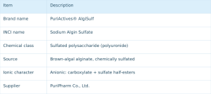
Distinct from unmodified alginate (INCI: Algin / Sodium Alginate), which contains no sulfate groups.
Source: Arlov & Skjåk-Bræk. Molecules 2017;22:778.
03 · MOLECULAR STRUCTURE
The alginate backbone and what sulfation changes
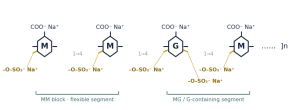
Schematic: (1→4)-linked β-D-mannuronic acid (M) and α-L-guluronic acid (G) residues. Gold bonds indicate sulfate half-esters at C2/C3 hydroxyl groups.
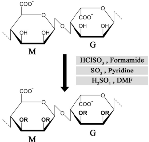
Published figure: chemical sulfation routes for alginate M/G residues (HClSO₃/formamide; SO₃/pyridine; H₂SO₄/DMF). Arlov & Skjåk-Bræk. Molecules 2017;22:778 (CC BY).
BACKBONE STRUCTURE
Alginate is a linear copolymer of M and G units arranged in MM, GG and alternating MG blocks. Block composition affects chain flexibility and ion binding. G blocks associate around Ca²⁺ to form the classic “egg-box” gel structure.
SULFATION CHEMISTRY
Chemical sulfation converts C2/C3 hydroxyl groups into sulfate half-esters (–O–SO₃⁻Na⁺), introducing additional anionic groups on the residues and producing a strongly acidic polyelectrolyte with a heparin-like spatial charge distribution.
Sources: Arlov & Skjåk-Bræk. Molecules 2017. DOI: 10.3390/molecules22050778; Mutch et al. 2024.
04 · STRUCTURE–ACTIVITY RELATIONSHIPS
Degree of sulfation shapes biological properties
Charge density influences protein affinity. Greater sulfation increases anionic charge along the chain and strengthens interactions with heparin-binding proteins, including growth factors, chemokines and coagulation factors (Biomacromolecules 2014).
Radical scavenging increases with sulfation. In human chondrocyte and macrophage models, superoxide scavenging by sulfated alginate correlates directly with the degree of substitution: more sulfate groups produce a stronger antioxidant response (Biomater. Sci. 2017).
Sequence and molecular weight regulate performance. M/G composition, block length and molecular weight shape chain conformation, flexibility and binding geometry. Sequence-defined samples show measurable differences in heparin-like behavior (Molecules 2017).
SODIUM ALGIN SULFATE VS UNMODIFIED SODIUM ALGINATE

Sources: Arlov et al. Biomacromolecules 2014; Kerschenmeyer et al. Biomater. Sci. 2017.
05 · MECHANISMS
Integrated mechanisms of action
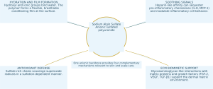
Sodium Algin Sulfate
Anionic sulfated polyuronide
HYDRATION AND FILM FORMATION
Hydroxyl and ionic groups bind water. The polymer forms a flexible, breathable conditioning film at the surface.
SOOTHING SIGNALS
Heparin-like affinity can sequester pro-inflammatory chemokines (IL-8, MCP-1) and modulate inflammatory cell behavior.
ANTIOXIDANT DEFENSE
Sulfate-rich chains scavenge superoxide radicals in a sulfation-dependent manner.
ECM BIOMIMETIC SUPPORT
Glycosaminoglycan-like interactions with matrix proteins and growth factors (FGF-2, VEGF, TGF-β1) support the dermal matrix environment.
One anionic backbone provides four complementary mechanisms relevant to skin and scalp care.
Sources: Huang et al. J. Nanobiotechnol. 2023; Freeman et al. Biomaterials 2008.
06 · SKIN APPLICATIONS: HYDRATION
Skin moisturization and conditioning
The structure of sulfated polyuronides supports water management. Dense hydroxyl, carboxylate and sulfate groups form hydrogen-bonding and ion–dipole interactions with water at the skin surface.
MOLECULAR WATER BINDING
Each residue carries hydroxyl groups, a carboxylate group and up to two sulfate half-esters. The source positions this as a high charge density among cosmetic polysaccharides, supporting hydrophilicity and moisture retention.
SURFACE FILM
Alginate materials have long been used in wound dressings for their ability to maintain a moist interface. As a topical film former, the polymer can reduce water loss and smooth surface roughness.
SENSORY CONDITIONING
Reviews of marine carbohydrates report moisturizing, soothing and anti-aging benefits of algal polysaccharides. Alginate-family materials are known for moisture-retaining protective films.
FORMULATION IMPLICATIONS
AlgiSulf can serve as a moisturizing framework in water-based serums, concentrates, gel masks and scalp treatments, providing hydration, slip and a soft-focus film without a heavy occlusive feel. Its anionic character also supports pairing with cationic conditioning complexes in rinse-off products.
Sources: Kim et al. Mar. Drugs 2018;16:459; Yao & Xu. Polysaccharides 2022.
07 · SKIN APPLICATIONS: BARRIER
Skin barrier support
Barrier care begins at the surface. A coherent, hydrated polymer film protects the compromised stratum corneum, while related chemical interactions may modulate inflammatory and oxidative signals that interfere with recovery.
Moist protective interface. Alginate dressings maintain a moist, protected wound environment. Sulfated alginate hydrogels extend this platform in deep partial-thickness burn models, reducing excessive inflammation and supporting organized repair (preclinical).
Fibroblast compatibility. Alginate sulfate showed no cytotoxicity in mouse and human fibroblasts at 0.5–2% w/v, while reducing inflammatory mediator levels in the same cells. This provides a rationale for barrier-care development for reactive skin (in vitro).
Bioadhesive residence. Alginate-family polymers exhibit mucoadhesion and bioadhesion, potentially extending surface film residence, a useful property for leave-on barrier products and overnight masks (material-class evidence).
Barrier support through signaling. By binding IL-8 and MCP-1 and promoting a shift from M1 toward M2 macrophage phenotypes, sulfated alginate acts upstream of low-grade inflammation associated with disrupted barrier homeostasis (preclinical).
Sources: Huang et al. J. Nanobiotechnol. 2023; Balde & Nazeer. Colloids Surf. B 2025.
08 · SKIN APPLICATIONS: SOOTHING
Soothing and inflammatory pathways
Sulfated alginate mimics heparan sulfate. Its negative charge distribution attracts positively charged chemokine domains, potentially intercepting signals before inflammatory cells are recruited into tissue.
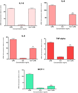
Published figure: unmodified sodium alginate from E. amentacea suppresses LPS-induced IL-1β, IL-6, IL-8, TNF-α and MCP-1 in THP-1 macrophages. Mar. Drugs 2026;24:41 (CC BY). DOI: 10.3390/md24010041.
CHEMOKINE SEQUESTRATION
● Sulfated alginate hydrogel platforms have high affinity for IL-8 and MCP-1 and reduce inflammatory cell infiltration (preclinical).
● Macrophage phenotypes shift from M1 toward M2, favoring resolution rather than escalation (preclinical).
INFLAMMATORY MEDIATOR MODULATION
● Alginate sulfate (0.5–2% w/v) reduces inflammatory mediators in mouse and human fibroblasts and inhibits protein denaturation (in vitro).
● Sulfated alginate hydrogels suppress IL-1β-induced inflammatory responses in human chondrocytes (in vitro).
BACKBONE EVIDENCE: UNMODIFIED SODIUM ALGINATE
For unmodified E. amentacea sodium alginate, reductions in THP-1 macrophages were IL-6: 46%; IL-8: 46%; MCP-1: 81%; TNF-α: 23%. iNOS inhibition in RAW 264.7 cells was 70–83%. These data describe the unsulfated backbone. The source separately proposes further enhancement through sulfation.
Sources: Huang 2023; Balde 2025; Arlov 2017; Mar. Drugs 2026;24:41.
09 · SKIN APPLICATIONS: ANTIOXIDANT ACTIVITY
Antioxidant protection
Reactive oxygen species contribute to skin aging and inflammation. Sulfation adds radical-scavenging activity to the alginate film platform, with activity increasing as sulfate content rises.
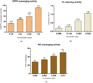
Published figure: DPPH scavenging (a), ferric reducing power (b) and NO scavenging (c) of unmodified E. amentacea sodium alginate. Mar. Drugs 2026;24:41 (CC BY). DOI: 10.3390/md24010041.
SULFATION-DEPENDENT SCAVENGING
Across sulfated alginate derivatives tested with human chondrocytes and macrophages, superoxide scavenging correlated directly with degree of sulfation, demonstrating a structure–activity relationship (in vitro).
The same study reported immunomodulatory effects, linking antioxidant activity with modulation of inflammatory signaling.
MARINE SULFATED POLYSACCHARIDES AS RADICAL SCAVENGERS
Sulfated polysaccharides from Laminaria japonica showed greater reducing power and radical scavenging at higher sulfate content and lower molecular weight. These are structural variables relevant to alginate sulfation (chemical assays).
QUANTITATIVE BACKBONE DATA: SODIUM ALGINATE
For the unsulfated backbone, DPPH scavenging reached 91% at 1.25 mg/mL (IC₅₀ ≈ 0.410 mg/mL). NO scavenging IC₅₀ was approximately 0.417 mg/mL. Ferric reducing power increased with dose to A₇₀₀ = 1.06. The source cites sulfation literature as a rationale for exceeding this baseline; the plotted data concern unsulfated alginate (chemical assays).
Sources: Kerschenmeyer 2017; Zhao et al. 2008; Mar. Drugs 2026;24:41.
10 · SKIN APPLICATIONS: AGING AND ECM
Skin aging and the extracellular matrix
Sulfated glycosaminoglycans participate in dermal matrix functions. Sulfated alginate uses related sulfate-patterned protein interactions and was originally developed for tissue repair applications.
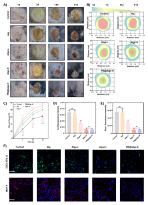
Published figure: in vivo closure of deep partial-thickness burn wounds over days 0–21 using a tunable sulfated alginate hydrogel platform. Huang et al. J. Nanobiotechnol. 2023;21:305 (CC BY). DOI: 10.1186/s12951-023-02144-2.
GROWTH FACTOR AFFINITY
Sulfated alginate hydrogels bind heparin-binding growth factors, including FGF-2, VEGF and TGF-β1, with high affinity and provide sustained release, mimicking the ECM’s storage of regenerative signals (in vitro).
REPAIR KINETICS
Connective tissue growth factor-loaded alginate sulfate nanoparticles achieved 79.49% wound closure at 72 hours with 5 µg/mL CTGF, the fastest among the tested groups (in vitro scratch assay).
ENZYME-RELATED PROTECTION: RELATED SULFATED POLYSACCHARIDES
Marine sulfated polysaccharides from green and red algae inhibit elastase, collagenase and hyaluronidase in vitro, enzymes relevant to wrinkle formation and loss of firmness. The source uses this material-class behavior as a rationale for AlgiSulf’s sulfate-rich surface.
Sources: Freeman 2008; Karam 2024; Huang 2023; Falcao 2020.
11 · EVIDENCE REVIEW
Key scientific evidence and quantitative findings

These studies used research-grade sulfated alginate or related sulfated polysaccharides. The findings characterize the material class and support the mechanistic rationale for PuriActives® AlgiSulf.
SCALP & HAIR SCIENCE
PART II · SCALP AND HAIR SCIENCE
The scalp is skin.
The hair shaft is keratin.
Hydration, soothing and antioxidant mechanisms relevant to facial skin also inform scalp microenvironment care, with further applications based on the electrostatics of the keratin hair surface.
12 · RATIONALE FOR SCALP CARE
The scientific rationale for scalp care
The scalp has dense follicles, greater sebum production and a resident microbiota, all relevant to hair condition. Common concerns, including tightness, itching, flaking and oiliness, involve hydration, barrier function and inflammation, areas relevant to sulfated polysaccharide research.
SCALP STRESSORS
● Sebum oxidation and UV exposure create oxidative stress in scalp skin and hair.
● Malassezia metabolites, such as oleic acid, disrupt barrier homeostasis and promote flaking.
● Surfactant cleansing and styling remove surface lipids and water.
WHERE ALGISULF FITS
● A moisturizing film addresses scalp dehydration and tightness.
● Chemokine affinity provides a rationale for targeting the itch–inflammation axis.
● Sulfation-related radical scavenging may buffer oxidative stress.
● A non-biocidal polymer approach is positioned for ongoing scalp care.
PATENT PRECEDENT
A patent claims sulfated polysaccharides as non-irritating anti-dandruff actives to prevent Malassezia-associated excessive flaking. Reconstructed skin data show complete suppression of oleic-acid-induced desquamation (EP2794012B1).
This provides a cosmetic application precedent for sulfated polysaccharides in scalp care.

Patent figure: the first page of EP2794012B1, claiming sulfated polysaccharides as cosmetic anti-dandruff actives (international classifications A61Q 5/00 and A61K 8/73).
Sources: Trüeb. Int. J. Trichology 2009; EP2794012B1; DeAngelis et al. 2005.
13 · SCALP HYDRATION
Scalp hydration and comfort
A dehydrated scalp can feel tight or itchy and show fine flaking after frequent washing. AlgiSulf’s polymer-based rationale is to bind water, maintain a film and modulate associated signals.
A surface water reservoir. The sulfate–carboxylate–hydroxyl network binds water without relying on heavy emollients, an important consideration for scalp products that should not weigh down hair.
Comfort through chemokine modulation. IL-8 and MCP-1 help recruit inflammatory cells in irritated skin. Demonstrated sulfated alginate affinity for these chemokines provides a mechanistic route relevant to scalp comfort (preclinical).
A non-stripping approach to flaking. In a patent model, sulfated polysaccharides prevented oleic-acid-induced desquamation in reconstructed skin, reducing the score from 3 to 0. The approach addresses flaking pathways rather than killing scalp microorganisms (patent, ex vivo).
Gentle wash-day feel. A smooth anionic film can reduce friction during massage and rinsing, supporting comfort in shampoos, scalp treatments and pre-wash products for sensitive scalps.
Sources: Huang et al. 2023; EP2794012B1.
14 · SCALP MICROENVIRONMENT
Scalp oxidative stress and the microenvironment
Oxidative stress is a documented contributor to scalp and hair aging. UV exposure and sebum lipid peroxidation increase ROS at the follicular environment, affecting scalp condition and the quality of emerging hair.
THE SCALP OXIDATIVE CASCADE

UV + SEBUM
Light exposure and sebum peroxidation generate ROS at the scalp surface and follicular openings.
OXIDATIVE STRESS
ROS irritate scalp skin, contribute to microinflammation and oxidize hair surface lipids.
VISIBLE EFFECTS
Scalp sensitivity, dull hair and progressive loss of hair quality.
ALGISULF BUFFERING CONCEPT
Sulfation-associated radical scavenging is proposed to interrupt the cascade at the surface.
MICROBIOME-FRIENDLY POSITIONING
Dandruff-associated dysbiosis involves Malassezia and Staphylococcus overgrowth. The source describes recurrence after stopping conventional antifungal treatment. Sulfated polysaccharides offer a proposed complementary, non-biocidal pathway: limiting metabolite-triggered flaking while preserving the resident ecosystem.
Sources: Trüeb. Int. J. Trichology 2009; Kerschenmeyer 2017; EP2794012B1.
15 · HAIR SHAFT
Hair conditioning through electrostatic interactions
Hair keratin has a net negative charge at cosmetic pH. Anionic polysaccharides can reach the hair surface through association with cationic conditioning polymers, which promote deposition and retention.
COMPLEXATION IMPROVES SUBSTANTIVITY
Complexing anionic glycosaminoglycan-like polysaccharides with cationic cellulose (polyquaternium-type polymers) increased keratin-bound polymer after rinsing by up to tenfold, with zeta-potential evidence of deposition (in vitro, hair tresses). The source proposes the same architecture for anionic sulfated polyuronides.
FILM-BASED CONDITIONING
After deposition, the sulfated film provides the conditioning functions associated with hair-care polysaccharides: cuticle smoothing, improved combability, moisture retention and slip, without the heavy feel associated with silicone oils.
The water-binding network helps maintain hair hydration between washes.
FORMULATION TIP
Pair AlgiSulf with cationic conditioners in conditioners and hair masks to engineer deposition. In leave-on scalp serums, the anionic film may function independently. One ingredient supports two delivery approaches.
Source: Pavlichko et al. Int. J. Cosmet. Sci. 1994; PMID 19250478.
16 · SYNERGY
Skin–scalp synergy: one INCI across facial, scalp and hair care
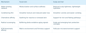
For brand developers, this alignment supports a consistent ingredient story across facial care, scalp care and hair care product lines.
17 · TECHNICAL PROPERTIES
Documented technical properties
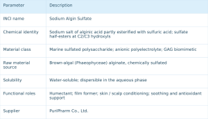
Full specifications, including appearance, content, viscosity and microbial limits, are provided in the product COA/TDS and can be requested from PuriPharm technical support. The spectra shown characterize the alginate backbone in a related material.
Published figure: FTIR fingerprint of the brown-algal sodium alginate backbone. Mar. Drugs 2026;24:41 (CC BY). DOI: 10.3390/md24010041.
Published figure: ¹H NMR anomeric-region spectrum used to resolve alginate M/G composition (same source). The presentation states that comparable COA-level characterization supports each AlgiSulf batch.
18 · FORMULATION
Formulation compatibility and processing
The following guidance is based on established polyelectrolyte chemistry of alginate-family polymers. Confirm specific parameters in pilot batches with PuriPharm technical support.
COMPATIBLE SYSTEMS
● Water-based and hydrogel systems: serums, concentrates, masks and scalp treatments.
● Nonionic and anionic ingredients: polyols, humectants and anionic surfactant bases.
● Oil-in-water emulsions: add to the aqueous phase for a light structural contribution.
● Cationic conditioners, when deliberately designed: controlled complexation for hair deposition.
● Near-neutral pH: a typical range for leave-on skin and scalp products.
HANDLE WITH CARE
● Strongly cationic ingredients: spontaneous polyelectrolyte complexation can be useful in hair care but should be avoided in clear gels.
● Low pH: uronic-acid protonation reduces charge and solubility; avoid strongly acidic systems.
● High electrolyte loading: charge screening can reduce viscosity and film quality.
● Multivalent cations, such as Ca²⁺: G-block “egg-box” gelation can be used deliberately as a texture tool.
PROCESSING
Add to the aqueous phase under moderate stirring and hydrate fully. A pre-dissolved solution may suit cold processing. Use levels depend on formulation goals; contact PuriPharm technical support.
19 · APPLICATIONS: SKINCARE
Skincare formulation opportunities
MOISTURIZING SERUMS / CONCENTRATES
A water-binding polyuronide film for a hydrated, plump feel with non-sticky slip.
SOOTHING GELS FOR SENSITIVE SKIN
Chemokine-affinity positioning for reactive, redness-prone or stressed skin in a lightweight hydrogel format.
BARRIER REPAIR CREAMS
Film-forming support inspired by dressings for compromised-barrier care and overnight repair masks.
ANTIOXIDANT DAY CARE
Sulfation-related radical scavenging as a concept for daily urban and photo-oxidative stress care.
FIRMING / ECM CARE CREAMS
GAG biomimetic positioning for matrix-care concepts in anti-aging and firming products.
SHEET MASKS AND ESSENCE SYSTEMS
A marine polysaccharide story with immediate surface smoothing and a soft after-feel.
CLAIM FRAMEWORK
Hydration · Barrier comfort · Soothing · Antioxidant defense · Matrix support. Each concept links to the mechanisms reviewed above.
20 · APPLICATIONS: SCALP AND HAIR
Hair and scalp formulation opportunities
SOOTHING SCALP SERUMS / TREATMENTS
Leave-on aqueous formats address tightness and itching through hydration and chemokine-affinity concepts.
FLAKING-CONTROL SCALP TREATMENTS
Patent precedent for sulfated polysaccharides supports a non-biocidal approach to flaking in daily scalp care.
SCALP HYDRATION MASKS
Pre-wash or overnight hydration for dry, tight scalps, with a light lotion texture and fresh feel.
CONDITIONERS / HAIR MASKS
Cationic complexation and deposition for cuticle smoothing, combability and sustained hair hydration.
AFTER-SUN SCALP SPRAYS
Antioxidant and soothing support for UV-exposed scalp partings, positioned as an emerging product opportunity.
GENTLE DAILY SHAMPOOS
Compatibility with anionic surfactants adds slip and post-wash comfort to mild, sulfate-free cleansing systems.
CLAIM FRAMEWORK
Scalp hydration and comfort · Flaking-control support · Oxidative stress defense · Hair conditioning.
Sources: EP2794012B1; Pavlichko et al. 1994; Trüeb 2009.
21 · EVIDENCE LANDSCAPE
Scientific evidence and application matrix
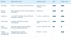
●●● Primary application area · ●● Strong supporting fit · ● Emerging rationale. These symbols reflect the depth of public evidence reviewed in the source presentation.
22 · VALUE PROPOSITION
What differentiates AlgiSulf
Beyond sodium alginate. Sulfation transforms a commodity texture polymer into a material with biological affinity, higher charge density, chemokine interactions and radical-scavenging properties, with a distinct INCI identity.
Characterized heparin-like chemistry. Two decades of peer-reviewed work on sulfated alginate, covering synthesis, sequence characterization, protein binding and cellular models, provide a substantial mechanistic literature for formulation concepts.
Continuity from skin to scalp. One ingredient carries a consistent marine polysaccharide story across facial, scalp and hair products, simplifying ingredient selection, claims development and supply chains.
Marine sourcing with contemporary relevance. Renewable brown-algal biomass, biodegradable polymer chemistry and a non-biocidal scalp-care concept align with responsible formulation goals.
PuriActives® AlgiSulf: marine sulfation science for formulation development.
23 · CONCLUSION AND REFERENCES
PuriActives® AlgiSulf: ready for your next formulation
A marine sulfated polysaccharide with a defined INCI identity, mechanistic literature and a combined skin–scalp application rationale. Technical support, samples and documentation are available from PuriPharm.
SELECTED REFERENCES
1. Arlov Ø, Skjåk-Bræk G. Sulfated alginates as heparin analogues: chemical and functional properties. Molecules 2017, 22(5), 778. DOI 10.3390/molecules22050778
2. Arlov Ø et al. Heparin-like properties of sulfated alginates with defined sequences and sulfation degrees. Biomacromolecules 2014, 15(7). DOI 10.1021/bm500602w
3. Kerschenmeyer A et al. Anti-oxidant and immune-modulatory properties of sulfated alginate derivatives on human chondrocytes and macrophages. Biomater. Sci. 2017, 5, 1756–1764. DOI 10.1039/C7BM00341B
4. Freeman I, Kedem A, Cohen S. Sulfation of alginate hydrogels: specific binding and controlled release of heparin-binding proteins. Biomaterials 2008, 29(22).
5. Huang C et al. Tunable sulfated alginate-based hydrogel platform for burn wound repair. J. Nanobiotechnology 2023, 21, 305. DOI 10.1186/s12951-023-02144-2
6. Balde A, Nazeer RA. In vitro anti-inflammatory effects of alginate sulfate; AlgS/PVA/CA microneedle system. Colloids Surf. B Biointerfaces 2025.
7. Karam M et al. Alginate and alginate sulfate/PCL nanoparticles for growth-factor delivery in wound healing. Biomed. Pharmacother. 2024.
8. Zhao X, Xue CH, Li BF. Antioxidant activities of sulfated polysaccharides from Laminaria japonica. J. Appl. Phycol. 2008. DOI 10.1007/s10811-007-9282-4
9. Mutch AL et al. Sulfated alginate for biomedical applications. Macromol. Biosci. 2024. DOI 10.1002/mabi.202400237
10. Kim JH et al. Beneficial effects of marine algae-derived carbohydrates for skin health. Mar. Drugs 2018, 16(11), 459. DOI 10.3390/md16110459
11. Ngo DH, Kim SK. Sulfated polysaccharides as bioactive agents from marine algae. Int. J. Biol. Macromol. 2013, 62.
12. Trüeb RM. Oxidative stress in ageing of hair. Int. J. Trichology 2009, 1(1), 6–14.
13. Pavlichko JP et al. Enhanced substantivity of hyaluronic acid on keratin substrates via polymer complexation. Int. J. Cosmet. Sci. 1994, 16(6). PMID 19250478
14. Use of sulphated polysaccharides as antidandruff agent. EP2794012B1 / WO2013093307A1 · Sodium alginate from E. amentacea: characterization & anti-inflammatory profile. Mar. Drugs 2026, 24(1), 41. DOI 10.3390/md24010041
PURIPHARM CO., LTD.
www.puriactives.com · service@puripharm.com · +86-572-2745768
Building 6, No. 1366 Hongfeng Road, Huzhou, Zhejiang, China
PuriActives® is a registered trademark of PuriPharm Co., Ltd.
NanoActive™ Harpa
NanoActive™ HarpaLiposomal Devil’s Claw Phytocomplex for Skin & Scalp Resilience
BRAND & PRODUCT
NanoActive™ Harpa · Liposomal Harpagoside Botanical Complex
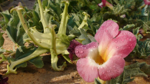
Liposomal Devil’s Claw Phytocomplex for Skin & Scalp Resilience
Powered by PURISOME® FlexVes™ Liposome Technology
Soothe inflammatory signals, restore barrier resilience and revitalize the scalp
PuriPharm Co., Ltd.
PRODUCT OVERVIEW
African desert botanical resilience meets modern nano-delivery
TRADE NAME
NanoActive™ Harpa
PRODUCT POSITIONING
Liposomal harpagoside botanical complex
INCI NAME
HARPAGOPHYTUM PROCUMBENS ROOT EXTRACT
BOTANICAL SOURCE
Devil’s claw root
MARKER ACTIVES
Harpagoside and other iridoid glycosides
DELIVERY TECHNOLOGY
PURISOME® FlexVes™ flexible vesicle liposome technology
APPLICATIONS
Skincare · Scalp care · Hair care
KEY BENEFITS
▪ Soothing and redness care
▪ Barrier recovery
▪ Reduced water loss
▪ Inflamm-aging care
▪ Scalp comfort
▪ Root vitality
PRODUCT VALUE
Botanical actives
FlexVes™ flexible liposomes
Skin and scalp applications
NanoActive™ Harpa
MARKET INSIGHT
Skin and scalp face shared inflammatory stress
DAILY STRESSORS
▪ Air pollution
▪ UV exposure and heat
▪ Extreme temperature changes
▪ Frequent cleansing
▪ Surfactant irritation
▪ Cosmetic procedures
▪ Shaving
▪ Hair coloring, bleaching and perming
▪ Heat styling
▪ Sleep and lifestyle stress
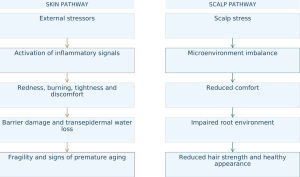
BOTANICAL ORIGIN
Devil’s claw: resilience from the African desert
Harpagophytum procumbens grows mainly in arid southern Africa, including Namibia, Botswana, South Africa, Angola and parts of Zimbabwe.
“Harpago” derives from the Latin word for “hook.” The fruit’s sharp hooks give the plant its common name, devil’s claw.
Adapted to extreme dryness: defensive secondary metabolites accumulate under desert heat and water stress.
Secondary storage roots are the traditional plant material used and contain concentrated actives.
Burgundy flowers and hooked fruits create a distinctive visual identity for the African desert botanical story.
Traditional use and scientific interest connect southern African botanical practice with modern inflammation research.

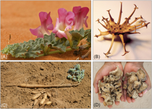
Namib Desert landscape; devil’s claw flowers, hooked fruits, secondary roots and dried roots (reference images).
Source: Zuplex Botanicals. Rosacalm Product Brochure. 2019 [5].
PHYTOCHEMICAL PROFILE
Beyond harpagoside: a multi-active botanical matrix
IRIDOID GLYCOSIDES
- Harpagoside (marker compound)
- Harpagide
- Procumbide
PHENOLIC GLYCOSIDES
- Acteoside / Verbascoside
- Isoacteoside
PHYTOSTEROLS
- β-Sitosterol
- TRITERPENES
- Oleanolic acid
- Ursolic acid
FLAVONOIDS
- Luteolin
- Kaempferol
AROMATIC ACIDS
- Caffeic acid
- Cinnamic acid
- Chlorogenic acid
Harpagoside is the principal marker compound. The full devil’s claw root extract also contains multiple complementary actives.
Source: Zuplex Botanicals. Rosacalm Product Brochure. 2019 [5].
FORMULATION PHILOSOPHY
One marker compound, multiple complementary pathways
SINGLE MARKER COMPOUND: HARPAGOSIDE
▪ A defined active marker
▪ A focus of mechanistic research
▪ A clear anchor for scientific communication
FULL DEVIL’S CLAW ROOT PHYTOCOMPLEX
▪ Multiple iridoid glycosides
▪ Complementary polyphenols and flavonoids
▪ Additional triterpenes
▪ Botanical activity across multiple targets
▪ A broader rationale for soothing and repair
NanoActive™ Harpa delivers a botanical active network centered on harpagoside.
Source: Abdelouahab N, Heard C. J Nat Prod. 2008;71:746–749. DOI: 10.1021/np070204u [2].
THE DELIVERY CHALLENGE
Botanical actives need effective delivery
Actives vary in polarity, making a single solvent system difficult to optimize.
Uneven partitioning between aqueous and oil phases affects formulation uniformity.
Suboptimal dispersion can limit active availability.
Limited skin or scalp contact time reduces surface residence.
Some actives are sensitive to light, oxidation and other environmental factors.
Conventional extracts offer limited technological differentiation for premium positioning.
Scalp actives may run off with liquid, further reducing contact efficiency.
PURISOME® FlexVes™ upgrades conventional devil’s claw extract into a functional active with a modern delivery concept.
DELIVERY TECHNOLOGY
Flexible vesicles improve contact with target surfaces
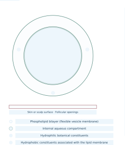
TECHNOLOGICAL DIFFERENTIATION
From conventional botanical extract to NanoActive™ delivery
| Dimension | Conventional devil’s claw root extract | NanoActive™ Harpa |
|---|---|---|
| Botanical source | Devil’s claw root | Active devil’s claw root phytocomplex |
| Marker constituent | Harpagoside | Multi-component phytocomplex centered on harpagoside |
| Dispersion | Conventional solution or dispersion | Phospholipid flexible vesicle delivery |
| Surface contact | Passive contact | Designed to enhance contact with skin and scalp surfaces |
| Technology story | Traditional African botanical | African botanical resilience + modern liposome technology |
| Applications | Primarily skin soothing | Skin, scalp and hair care platform |
| Market positioning | Natural botanical extract | Premium botanical active with nano-delivery |
MECHANISM OVERVIEW
Soothing begins with upstream signaling
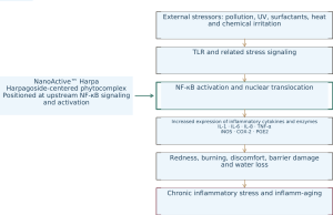
KEY MESSAGE
Modulation of upstream NF-κB-related signals provides a mechanistic rationale for soothing, redness care and barrier repair with devil’s claw root actives.
Source: Huang THW et al. J Ethnopharmacol. 2006;104:149–155. DOI: 10.1016/j.jep.2005.08.055 [1].
MECHANISM: NF-κB
NF-κB: a key regulator of inflammatory cascades
NF-κB links external stimuli with inflammatory mediator expression.
After stimulation, NF-κB can activate and enter the nucleus, promoting inflammatory mediators associated with redness, heat sensations and barrier discomfort.
Studies of harpagoside report effects on NF-κB activation and nuclear translocation, providing a molecular rationale for upstream inflammatory signal management.
The mechanistic evidence comes from published cellular and molecular studies and supports the product’s soothing rationale [1].
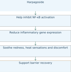
Source: Huang THW et al. J Ethnopharmacol. 2006;104:149–155 [1].
MECHANISM: INFLAMMATORY MEDIATORS
Inflammatory mediator regulation at multiple levels
iNOS
Inducible nitric oxide synthase
▪ Involved in NO production during inflammation
▪ Excess activity is associated with oxidative stress
▪ May increase cellular stress
COX-2
Inducible cyclooxygenase
▪ An important inducible enzyme in inflammation
▪ Participates in prostaglandin production
▪ Associated with redness, burning and discomfort
PGE2
Prostaglandin E2
▪ Involved in local vasodilation and inflammatory signaling
▪ May contribute to redness, swelling and barrier discomfort
NANOACTIVE™ HARPA: MECHANISTIC POSITIONING
NF-κB — iNOS — COX-2 — PGE2 inflammatory signaling axis
Harpagoside and the full phytocomplex provide a rationale for soothing at multiple levels of this axis.
Sources: Huang THW et al., 2006 [1]; Abdelouahab N, Heard C., 2008 [2].
SKIN INTERFACE RESEARCH
From cellular signaling to topical skin applications
▪ Devil’s claw root extract and its major glycosides have been studied in skin-related models [2].
▪ Research includes epidermal COX-2 responses relevant to redness and discomfort signals.
▪ Constituent ratios may influence overall effects; a single compound may not substitute for the full extract.
▪ Skin models indicate complementary value from multiple constituents.
▪ Transcutaneous and topical studies provide a mechanistic basis for cosmetic soothing applications [3].
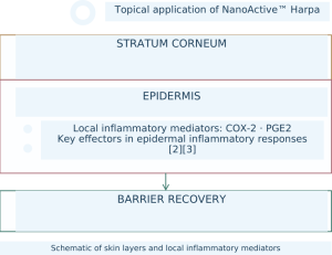
Skin model research connects molecular mechanisms with topical use, supporting the rationale for soothing and barrier repair.
Sources: Abdelouahab N, Heard C. J Nat Prod. 2008 [2]; Int J Pharm. 2009 (ex vivo skin model study) [3].
INFLAMM-AGING
Managing low-grade inflammatory stress for skin resilience
Persistent low-grade inflammatory stress may affect skin cells, barrier function and the extracellular matrix. Inflamm-aging is therefore a concept used in premium anti-aging product development.

APPLICATION TERRITORIES
▪ Anti-aging for sensitive skin
▪ Repair-focused anti-aging
▪ Urban environmental care
▪ Barrier-focused anti-aging
▪ Soothing support alongside potent actives
▪ Skin longevity and resilience concepts
Upstream signal management provides a scientific rationale for combining soothing with anti-aging product concepts.
Mechanistic sources: Huang THW et al., 2006 [1]; Abdelouahab N, Heard C., 2008 [2].
EFFICACY BENCHMARK: THIRD-PARTY DEVIL’S CLAW ROOT EXTRACT DATA
Redness and water loss improvements over 24–48 hours
TEST DESIGN
TEST LABORATORY:Dermaconsult GmbH
PARTICIPANTS:20 participants, plus 1 reserve participant
SEX AND AGE:14 women and 6 men; 23–61 years
TEST AREA:Inner forearm
ACTIVE CONCENTRATION:5%
ASSESSMENTS:Skin redness · Transepidermal water loss · Skin irritation patch test
INSTRUMENTS:CR-400 chroma meter · Tewameter Triple TM 330 T
INSTRUMENTAL RESULTS
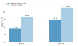
| Endpoint | 24 hours | 48 hours |
|---|---|---|
| Reduction in skin redness | 9.1% | 14.7% |
| Reduction in TEWL | 16.7% | 22.95% |
▪ Redness improvement observed at 24 hours
▪ Greater improvement at 48 hours
▪ Continued reduction in transepidermal water loss
▪ Supports positioning for rapid soothing and barrier recovery
▪ Relevant to products for sensitive, irritated or environmentally stressed skin
Note: These data come from a third-party human efficacy test of 5% conventional devil’s claw root extract. They serve as botanical benchmarks for NanoActive™ Harpa product development and efficacy positioning.
Sources: Zuplex Botanicals. Rosacalm Product Brochure. 2019 [5]; Dermaconsult GmbH Test Summary [6].
SKINCARE PLATFORM
From rapid soothing to lasting barrier resilience
CALM
▪ Help reduce visible redness
▪ Soothe tightness and discomfort
▪ Help restore skin comfort
RESTORE
▪ Support barrier recovery
▪ Help reduce transepidermal water loss
▪ Improve dryness, roughness and fragility
DEFEND
▪ Help manage inflammatory signals associated with external stressors
▪ Support care under pollution and climate stress
▪ Provide soothing support in formulations with potent actives
REVITALIZE
▪ Help restore a healthy, balanced appearance
▪ Improve softness and moisturized feel
▪ Support repair-focused anti-aging and skin resilience concepts
Rapid botanical soothing supports skin recovery and resilience.
SKINCARE APPLICATIONS
For repair-focused and sensitive-skin products
01 Redness rescue serum
Rapid soothing care for environmental irritation, temperature changes, sleep loss and fragile skin.
02 Urban repair cream
For skin stressed by pollution, dryness and demanding lifestyles.
03 Soothing serum to complement potent actives
For concepts featuring retinol, retinal, acids or high-concentration niacinamide.
04 After-sun soothing lotion
Comfort care following UV and heat exposure.
05 Eye-area soothing serum
For the appearance of fatigue, dryness and puffiness.
06 Post-shave barrier repair lotion
For redness, tightness and discomfort after shaving.
OTHER APPLICATIONS
Soothing masks · Body repair lotions · Hand and foot care · Cosmetic aftercare for tattoos · Local repair gels
COMPLEMENTARY INGREDIENT CONCEPTS
Panthenol · Ectoin · β-Glucan · Niacinamide · Ceramides · Bisabolol · Allantoin · Hyaluronic acid
SCALP SCIENCE
Scalpification brings barrier and soothing science to the scalp
SCALP STRESSORS
▪ Frequent shampooing
▪ Surfactants
▪ Sebum oxidation
▪ Pollutant accumulation
▪ UV and heat
▪ Hair coloring, bleaching and perming
▪ Blow-drying and heat styling
▪ Scratching and mechanical friction
POTENTIAL IMPACT
▪ Tightness and discomfort
▪ Visible redness
▪ Barrier imbalance
▪ Dryness or sebum imbalance
▪ Affected root environment
▪ Reduced hair strength and healthy appearance
Close-up scalp and hair-root images (reference images).
NanoActive™ Harpa extends sensitive-skin soothing science to scalp microenvironment care.
HAIR CARE DIRECTION: PATENT-SUPPORTED APPLICATION CONCEPTS
From botanical soothing to root vitality
HENKEL AG & CO. KGAA
Patent WO2008031685A1
Root Extract of Harpagophytum for Stimulating Hair Growth
Published patent concerning devil’s claw root extract for hair applications [4].
▪ Devil’s claw actives appear in hair strengthening and thickening concepts.
▪ The patent addresses root vitality and hair-condition improvement.
▪ Applications include shampoos, conditioners, scalp serums, sprays, lotions and scalp tonics.
▪ Patent examples include formulations with devil’s claw extract.
▪ Studies involve dermal papilla cells and keratinocytes.
▪ The patent discusses advantages of liposomal encapsulation in scalp and hair care products.
These patent-supported development directions provide technical and conceptual references for NanoActive™ Harpa scalp and hair care applications.
Source: Henkel AG & Co. KGaA. Patent WO2008031685A1 [4].
SCALP DELIVERY
Improving botanical contact with scalp and root surfaces
CONVENTIONAL WATER-SOLUBLE BOTANICAL EXTRACT
▪ Can run off with liquid
▪ Limited surface residence
▪ Potentially uneven distribution
▪ Shorter scalp contact time
NanoActive™ Harpa
FLEXVES™ LIPOSOMAL DELIVERY
▪ Delivery associated with phospholipid vesicles
▪ Improved scalp surface distribution
▪ Enhanced contact with follicular openings
▪ Suitable for leave-on scalp serums and tonics
▪ A modern technology concept for root care
▪ Integration of soothing and root vitality concepts
Liposomal delivery is designed to support retention, uniform distribution and longer contact on the scalp.
SCALP & HAIR PLATFORM
Scalp comfort, root strength and revitalized hair
SCALP COMFORT
▪ Help soothe tightness and discomfort
▪ Improve comfort in irritated scalps
▪ Support scalp barrier balance
ROOT VITALITY
▪ Support a healthy root microenvironment
▪ Suitable for volumizing, strengthening and anti-hair-loss product concepts
▪ Help improve the appearance of thinning and fragile hair
HAIR STRENGTH
▪ Support damaged and fragile hair care
▪ Help improve perceived strength and healthy shine
▪ Suitable for care after coloring and perming
MARKET DIFFERENTIATION
African devil’s claw botanical story · Harpagoside mechanisms · Skin and scalp applications · Nano-liposome technology · Soothing combined with root vitality
Scalp soothing is the starting point for a stronger, fuller-looking hair concept.
SCALP & HAIR PRODUCT CONCEPTS
From daily scalp balance to intensive root care
01 Scalp soothing serum
For tightness, dryness and scalp discomfort.
02 Root vitality serum
For volume, strength and healthy-root concepts.
03 Sensitive-scalp shampoo
Combines gentle cleansing with scalp soothing.
04 Root strengthening conditioner
Supports smoothness and a stronger hair appearance.
05 Scalp repair serum after chemical treatments
Comfort care after coloring, bleaching or perming.
06 Volumizing leave-on spray
Lightweight, non-sticky daily root care.
COMPLEMENTARY INGREDIENT CONCEPTS
Caffeine · Niacinamide · Panthenol · Zinc PCA · Amino acids · Peptides · Ceramides · Botanical antioxidants
NANOACTIVE™ HARPA
African desert resilience translated into beauty care
SOOTHE INFLAMMATORY SIGNALS
A multi-component botanical system centered on harpagoside supports modern skin and scalp soothing concepts.
RESTORE BARRIER RESILIENCE
Third-party instrumental testing of the conventional extract supports positioning for redness care, reduced water loss and barrier recovery.
REVITALIZE THE SCALP
Patent-supported root-care directions and PURISOME® flexible vesicle delivery expand the possibilities for premium scalp and hair products.
NanoActive™ Harpa combines devil’s claw botanical resilience with modern nano-liposome technology in an integrated concept for skin soothing, barrier recovery, scalp balance and root vitality.

NanoActive™ Harpa · Powered by PURISOME® FlexVes™ Liposome Technology
PURIPHARM CO., LTD.
REFERENCES
References and source materials
[1] Huang THW, Tran VH, Duke RK, et al. Harpagoside suppresses lipopolysaccharide-induced iNOS and COX-2 expression through inhibition of NF-kappa B activation. Journal of Ethnopharmacology. 2006;104(1–2):149–155. DOI: 10.1016/j.jep.2005.08.055
[2] Abdelouahab N, Heard C. Effect of the major glycosides of Harpagophytum procumbens on epidermal COX-2 in vitro. Journal of Natural Products. 2008;71(5):746–749. DOI: 10.1021/np070204u
[3] A Novel Ex Vivo Skin Model for the Assessment of the Potential Transcutaneous Anti-inflammatory Effect of Topically Applied Harpagophytum procumbens Extract. International Journal of Pharmaceutics. 2009.
[4] Henkel AG & Co. KGaA. Root Extract of Harpagophytum for Stimulating Hair Growth. Patent WO2008031685A1.
[5] Zuplex Botanicals. Rosacalm Product Brochure. 2019.
[6] Dermaconsult GmbH. Third-party instrumental evaluation of skin redness, transepidermal water loss and skin irritation, as summarized in the Rosacalm technical presentation.
[7] European Commission CosIng Database. HARPAGOPHYTUM PROCUMBENS ROOT EXTRACT.
NanoActive™ Oryzanol
01 · NanoActive™ Oryzanol: Oryzanol Nanoliposomes
Cosmetic active ingredient product introduction. INCI: ORYZANOL. Rice-derived antioxidant defense, reinvented by liposomal delivery.
Powered by PURISOME® FlexVes™ Liposome Technology. PuriPharm Co. Ltd.

02 · From Rice-Bran Active to Nano-Delivered Antioxidant
γ-Oryzanol is a group of phytosterol/triterpene alcohol ferulate esters naturally found in rice bran oil. Sterol lipids provide lipid affinity, while ferulate groups provide radical-defense potential.
Antioxidant: radicals/lipid peroxidation. Photoaging care: light-induced oxidative stress. Aging care: collagen/elastic-network concepts. Skin + Hair: skin, scalp, and hair.
NanoActive™ uses lipid vesicles to improve practical cosmetic formulation design.
03 · γ-Oryzanol Ingredient Science
A mixture of ferulate esters in the unsaponifiable fraction of rice bran oil. Representative components: Cycloartenyl Ferulate, 24-Methylene Cycloartanyl Ferulate, Campesteryl Ferulate, and Sitosteryl Ferulate.
An ester bond links the phenolic antioxidant ferulic-acid moiety with a lipid-affine phytosterol/triterpene-alcohol moiety.
Sources: Lerma-García et al. Food Chem. 2009;115:389–404; Ramazani et al. Curr Pharm Des. 2021;27(19):2299–2316. DOI: 10.2174/1381612826666201102101428.
Source link: https://doi.org/10.2174/1381612826666201102101428
References: Lerma-García JM, et al. Food Chemistry. 2009;115:389–404. | Ramazani E, et al. Curr Pharm Des. 2021;27(19):2299–2316. DOI: 10.2174/1381612826666201102101428
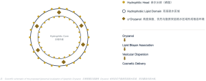
04 · Plant Antioxidant and Skin Lipid Science
Image: rice bran, the natural source.
01 Radical defense and oxidative-chain inhibition. 02 Lipid antioxidant activity in lipid-rich environments. 03 Ferulate UV-absorption/antioxidant properties as a basis for photostress care. 04 Research on collagenase, elastase, hyaluronidase, and tyrosinase.
05 · Multiple Antioxidant and Aging-Care Pathways
The mechanism map includes lower ROS, lipid peroxidation, oxidative damage, collagenase, elastase, hyaluronidase, and tyrosinase.
Concepts: antioxidant/photoaging care, elasticity, hydration-related structures, even tone, soothing defense, and scalp/hair antioxidants. These summarize in vitro/model literature and do not establish finished-product clinical efficacy.
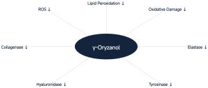
06 · Radical-Chain Management and Lipid Protection
Ferulate esters participate in radical scavenging and reduced radical-induced lipid peroxidation. Relevant lipid environments include stratum-corneum lipids, sebum, cellular membranes, hair surface lipids, and oxidizable formulation oils.
Pathway: radical scavenging, lipid protection, and biological-structure protection.
Juliano C, Cossu M, Alamanni MC, Piu L. Int J Pharm. 2005;299:146–154. DOI: 10.1016/j.ijpharm.2005.05.018.
Source link: https://doi.org/10.1016/j.ijpharm.2005.05.018
Juliano C, Cossu M, Alamanni MC, Piu L. Antioxidant activity of gamma-oryzanol: mechanism of action and its effect on oxidative stability of pharmaceutical oils. Int J Pharm. 2005;299:146–154. DOI: 10.1016/j.ijpharm.2005.05.018
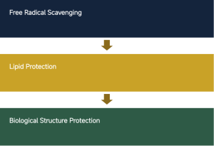
07 · Oxidative Stress Connects Exposure and Skin Aging
UV/pollution/ROS, oxidative stress, cellular damage, ECM degradation, and loss of elasticity/roughness/uneven tone form the schematic pathway.
NanoActive™ Oryzanol uses antioxidant design for daily aging-care concepts. This does not replace sunscreen and provides no SPF/PA claims.
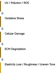
08 · 2026 Nanoencapsulated γ-Oryzanol Research
Literature nanoemulsion, H₂O₂-induced NIH/3T3 mouse fibroblast aging model: reduced intracellular ROS, supported survival/migration, and inhibition of collagenase, elastase, hyaluronidase, and tyrosinase.
Reported parameters: approximately 162 nm particle size, PDI 0.2, and zeta potential −51 mV. These are Yadav’s independent nanoemulsion values, not NanoActive™ product parameters.
Yadav P, Sankar J, Mora C, et al. Nanoencapsulated gamma-oryzanol enhances fibroblast proliferation, migration, and reduces oxidative stress in H₂O₂-induced cellular aging model. J Funct Foods. 2026;136:107130. DOI: 10.1016/j.jff.2025.107130.
Source link: https://doi.org/10.1016/j.jff.2025.107130
Yadav P, Sankar J, Mora C, et al. Nanoencapsulated gamma-oryzanol enhances fibroblast proliferation, migration, and reduces oxidative stress in H₂O₂-induced cellular aging model. J Funct Foods. 2026;136:107130. DOI: 10.1016/j.jff.2025.107130
09 · Protecting Collagen, Elastin, and Hyaluronic-Acid Networks
The dermal ECM schematic shows collagen, elastin, and hyaluronic acid. Oxidative stress and collagenase/elastase/hyaluronidase activity can contribute to network breakdown.
Multi-enzyme defense extends the literature concept from ROS management to structural-network preservation.
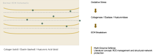
10 · Fibroblast Vitality
Dermal fibroblasts maintain collagen, elastin, and ECM. Oxidative pressure can reduce vitality/migration and accelerate structural decline.
Yadav’s 2026 nanoemulsion study reported ROS reduction, viability support, and migration in an oxidative model, informing cellular vitality, matrix defense, and resilience concepts.
J Funct Foods. 2026;136:107130. DOI: 10.1016/j.jff.2025.107130. Independent formulation evidence.
Source link: https://doi.org/10.1016/j.jff.2025.107130
Source: Yadav P, et al. J Funct Foods. 2026;136:107130 (literature nanoemulsion, not this product)。DOI: 10.1016/j.jff.2025.107130
11 · Tone and Brightening Research
2025 in vitro B16F10 mouse melanoma research: DPPH/FRAP antioxidant activity, lower cellular ROS, mushroom tyrosinase inhibition, reduced cellular melanin, and melanogenesis-related inhibition.
Concepts: antioxidant brightening, tyrosinase modulation, and even tone. In vitro potential does not establish clinical brightening or product efficacy.
Emami SA, Hadipour E, Boozari M, et al. Comparison of Antioxidant and Anti-Melanogenic Activity of γ-Oryzanol and Gallic Acid by Inhibiting Tyrosinase Activity in B16F10 Mouse Melanoma Cells. Food Sci Nutr. 2025;13:e70946. DOI: 10.1002/fsn3.70946.
Source link: https://doi.org/10.1002/fsn3.70946
Emami SA, Hadipour E, Boozari M, et al. Comparison of Antioxidant and Anti-Melanogenic Activity of γ-Oryzanol and Gallic Acid by Inhibiting Tyrosinase Activity in B16F10 Mouse Melanoma Cells. Food Sci Nutr. 2025;13:e70946. DOI: 10.1002/fsn3.70946
12 · UV-Associated Antioxidant Defense
Schematic: UV exposure, ROS generation, lipid peroxidation, inflammatory stress, and ECM damage.
Cosmetic language: helps defend against UV-induced oxidative stress; supports daily photostress protection; helps protect skin lipids from oxidative challenge.
No SPF/PA values, sunscreen replacement, or core regulatory claim of a natural sunscreen.

13 · Nanoethosome Delivery Evidence
2021 γ-oryzanol nanoethosomes improved delivery/cellular uptake, offered long-term storage protection, and showed protection in UVB-related models.
The original mouse work involved UV-induced skin cancer; this article retains it as delivery/photostress research, without disease-prevention/treatment claims.
Zeinali M, et al. Prevention of UV-induced skin cancer in mice by gamma oryzanol-loaded nanoethosomes. Life Sci. 2021;283:119759. DOI: 10.1016/j.lfs.2021.119759.
Source link: https://doi.org/10.1016/j.lfs.2021.119759
Zeinali M, et al. Prevention of UV-induced skin cancer in mice by gamma oryzanol-loaded nanoethosomes. Life Sciences. 2021;283:119759. DOI: 10.1016/j.lfs.2021.119759
14 · Formulation Challenges
Low water dispersibility; crystallization/precipitation with system or temperature changes; reliance on oils, emulsions, or solubilizers; limited effective delivery of free active; and prolonged environmental exposure.
Published approaches include solubilization, dispersion, emulsification, phospholipid complexes, and nanodelivery.
Patent examples: JP6803732B2, Cosmetics containing γ-oryzanol; WO2012110303A2, Aqueous cosmetic composition containing composite material particles and gamma-oryzanol; EP1957041B1, Oil-in-water emulsion and its use for delivery of functionality.
Patent examples (publication number and title): JP6803732B2 · Cosmetics containing γ-oryzanol;WO2012110303A2 · Aqueous cosmetic composition containing composite material particles and gamma-oryzanol;EP1957041B1 · Oil-in-water emulsion and its use for delivery of functionality。
15 · From Lipophilic Active to Formulation-Ready Nano Active
Oryzanol, PURISOME® FlexVes™, and NanoActive™ Oryzanol form the design pathway.
Protection: a phospholipid microenvironment. Dispersion: an adapted form for cosmetics. Delivery: effective skin-surface contact potential. Flexibility: serums, lotions, creams, scalp, and hair products.
The design changes incorporation and contact rather than merely adding free Oryzanol.

16 · Biomimetic Lipid Vesicle Architecture
The scientific schematic labels a hydrophilic aqueous core, phospholipid hydrophilic heads, and a hydrophobic bilayer domain. Highly lipophilic γ-Oryzanol is proposed to associate preferentially with the bilayer rather than the inner aqueous core.
Oryzanol, lipid-bilayer association, vesicular dispersion, and cosmetic delivery. The localization drawing is proposed architecture rather than a direct measurement.
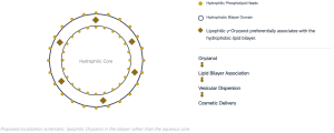
17 · More Than 15 Years of Liposome Research
2009 topical γ-oryzanol liposomes: phosphatidylcholine, cholesterol, CHAPS, and sodium taurocholate. Initial loading levels 3%, 5%, and 10%.
Endpoints: size, zeta potential, entrapment, release, antioxidant activity, and cellular compatibility. Antioxidant activity remained after loading; some systems showed enhanced cellular antioxidant activity.
Viriyaroj A, Ngawhirunpat T, Sukma M, et al. Physicochemical properties and antioxidant activity of gamma-oryzanol-loaded liposome formulations for topical use. Pharm Dev Technol. 2009;14(6):665–671. DOI: 10.3109/10837450902911937. Literature formulation, not product data.
Source link: https://doi.org/10.3109/10837450902911937
Viriyaroj A, Ngawhirunpat T, Sukma M, et al. Physicochemical properties and antioxidant activity of gamma-oryzanol-loaded liposome formulations for topical use. Pharm Dev Technol. 2009;14(6):665–671. DOI: 10.3109/10837450902911937 —literature formulation supporting NanoActive™ design, not product data.
18 · Research from Liposomes to Lipid Nanocarriers
2009 topical liposomes; 2009 solid lipid nanoparticles/nanoemulsions (SLN/NE); 2021 nanoethosomes; 2024 liposomal nanocarriers; 2026 cellular-aging nanoencapsulation.
Independent teams use these strategies for stability, dispersion, and delivery, providing continuity for NanoActive™ design.
Ruktanonchai U, et al. J Microencapsul. 2009;26(7):614–626. DOI: 10.3109/02652040802586571; Jasim AJ, et al. ACS Omega. 2024;9:3554–3564. DOI: 10.1021/acsomega.3c07339. Other sources appear in their sections.
Source link: https://doi.org/10.3109/02652040802586571
Source link: https://doi.org/10.1021/acsomega.3c07339
Ruktanonchai U, et al. J Microencapsul. 2009;26(7):614–626. DOI: 10.3109/02652040802586571 | Jasim AJ, et al. ACS Omega. 2024;9:3554–3564. DOI: 10.1021/acsomega.3c07339 (see other research sections)
19 · Human Evidence from Composite Rice-Bran Vesicles
Composite system: Ferulic Acid + γ-Oryzanol + Phytic Acid + Niosomes. 30 volunteers, 28 days.
Endpoints: hydration, elasticity, thickness, roughness, and pigmentation. Gels/creams containing the composite vesicles improved multiple aging-related indicators.
All human effects cannot be attributed to γ-oryzanol alone. The study supports composite rice-bran actives and vesicular delivery, not direct NanoActive™ efficacy.
Manosroi A, Chutoprapat R, Abe M, Manosroi W, Manosroi J. Pharm Biol. 2012;50(2):208–224. DOI: 10.3109/13880209.2011.596206.
Source link: https://doi.org/10.3109/13880209.2011.596206
Manosroi A, Chutoprapat R, Abe M, Manosroi W, Manosroi J. Anti-aging efficacy of topical formulations containing niosomes entrapped with rice bran bioactive compounds. Pharm Biol. 2012;50(2):208–224. DOI: 10.3109/13880209.2011.596206
20 · Antioxidant Defense and Inflammatory Stress
Non-skin models suggest effects on NF-κB-associated inflammatory signaling. These provide research clues for oxidative/inflammatory-stress care.
Cosmetic concepts: comfort support, environmental defense, and reduced oxidative burden in sensitive/tired/stressed skin. No medicinal anti-inflammatory or disease-treatment claims.
Ramazani E, Akaberi M, Emami SA, Tayarani-Najaran Z. Curr Pharm Des. 2021;27(19):2299–2316. DOI: 10.2174/1381612826666201102101428.
Source link: https://doi.org/10.2174/1381612826666201102101428
Ramazani E, Akaberi M, Emami SA, Tayarani-Najaran Z. Biological and Pharmacological Effects of Gamma-oryzanol: An Updated Review of the Molecular Mechanisms. Curr Pharm Des. 2021;27(19):2299–2316. DOI: 10.2174/1381612826666201102101428
21 · From Skin Lipids to Hair Lipids
Hair surface lipids face oxidative/environmental stress. Concepts: scalp antioxidant defense; hair lipid protection; sun/pollution/heat-styling care; and phospholipid conditioning support.
Patents: EP0945122A1, Haircare composition containing gamma-oryzanol and calcium salts; WO2012110303A2, skin/hair photoprotection context.
Claims stay within antioxidants, lipid care, and smoothness, excluding hair growth or hair-loss prevention.
EP0945122A1 — Haircare composition containing gamma-oryzanol and calcium salts WO2012110303A2 — relates to skin and/or hair photoprotection against ultraviolet radiation
22 · One Platform, Skin and Hair
Skin: antioxidants, photoaging-related stress, collagen/elastic matrix, hyaluronic-acid-related structures, tone, and environmental comfort.
Scalp/hair: antioxidants, fiber oxidation protection, photostress, lipids, and conditioning. Hair-growth/hair-loss claims are excluded.
23 · Modern Formulation Applications
Skin: antioxidant/aging/day-defense serums, photoaging-care lotion, recovery cream, brightening serum, urban protection, after-sun care, eye-aging care, and premium rice-bran concepts.
Scalp: antioxidant serum, comfort care, post-sun care, and urban-pollution care. Hair: conditioner, mask, leave-on serum, oil/emulsion, sun-exposure care, post-color/perming care, and heat-styling care.
The source cites commercial approximately 5% γ-oryzanol emulsion ingredients for soap/facial wash, lotion, shampoo, and conditioner as market context, not this product’s active content or recommended use level.
24 · A Formulation-Friendly Oryzanol Design
Free active challenges: hydrophobicity, poor aqueous dispersion, crystallization, oil/solubilizer dependence, complex processing, restricted formats, and delivery limitations.
NanoActive™ design: lipid-vesicle organization, aqueous dispersion, handling convenience, a lipid-compatible microenvironment, serum/emulsion use, and skin/hair applications.
This is qualitative dosage-form/process comparison, without unverified fold-improvement claims. Actual performance requires formulation validation.
25 · Rice Antioxidant, Reinvented
Rice-derived science; radical/lipid-peroxidation defense; ECM protection concepts; antioxidant tone support; skin/scalp/hair use; and PURISOME® FlexVes™ liposomal delivery.
NanoActive™ Oryzanol brings a classic rice-bran antioxidant into precision lipid-delivery design.
PuriActives® SLL
01 · PuriActives® SLL: Sodium Lauroyl Lactylate
Technical product presentation. INCI: SODIUM LAUROYL LACTYLATE. Formula C18H31NaO6, MW approximately 366.4 g/mol.
Multifunctional lactylate interface technology for skin and hair care. More Than an Emulsifier. PuriPharm Co., Ltd.

02 · More Than an Emulsifier
Positioning: a multifunctional interface ingredient.
01 Emulsification: uniform oil/water dispersion and O/W interface stability. 02 Mild cleansing: an anionic structure supporting mild-system design. 03 Lipid compatibility: a C12 chain and lactylated headgroup for complex lipid formulations. 04 Skin/hair sensory properties: softer, refined cleansing and care experiences.
Its amphiphilic structure participates in oil/water interfaces, surfactant assemblies, and lipid formulations across skin and hair applications.
03 · From Strong Cleansing to Cleansing Quality
Traditional focus: strong cleansing, high foam, and simple emulsions. Modern needs: mild cleansing, barrier-conscious design, lipid compatibility, refined sensory properties, multifunctionality, and skin/scalp-friendly concepts.
SLL offers an interface-chemistry approach to these formulation needs.
04 · Product Identity
The product information table and molecular structure are retained below.
Class: acyl lactylate, anionic surfactant. CosIng function: Surfactant–Emulsifying.
Sources in the presentation: PubChem—Sodium Lauroyl Lactylate; EU CosIng; Johnson et al. Int J Toxicol. 2024.
Source link: https://pubchem.ncbi.nlm.nih.gov/compound/Sodium-lauroyl-lactylate
Source link: https://ec.europa.eu/growth/tools-databases/cosing/
| Item | Information |
|---|---|
| Product | PuriActives® SLL |
| INCI | SODIUM LAUROYL LACTYLATE |
| Chinese INCI | 月桂酰乳酰乳酸钠 (Sodium Lauroyl Lactylate) |
| CAS No. | 13557-75-0 |
| EC No. | 236-942-6 |
| Molecular Formula | C18H31NaO6 |
| Molecular Weight | Approximately 366.4 g/mol |
| Ingredient Class | Acyl Lactylate · Anionic Surfactant |
| CosIng Function | Surfactant – Emulsifying |
05 · Fatty Acid × Lactic Acid Chemistry
Lauric acid, a C12 fatty acid, combines with lactic-acid/lactylate chemistry. The schematic shows esterification followed by neutralization to the sodium salt.
Fatty-acid-derived lipophilicity and lactylate-derived hydrophilicity produce a balanced amphiphilic architecture: a molecular bridge between water and lipids.
06 · How SLL Works at the Interface
The hydrophilic lactylated head faces water; the hydrophobic C12 lauroyl chain enters the oil/lipid phase.
01 Adsorption: molecules migrate to the oil/water interface. 02 Organization: the chain anchors in oil and the head faces water. 03 Dispersion support: interface organization provides a structural basis for stability.
This amphiphilic architecture supports emulsions, cleansing systems, and lipid formulations.
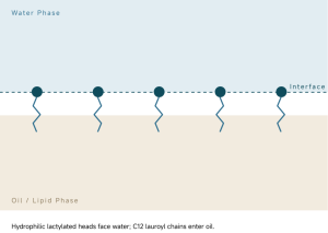
07 · Emulsification and Interface Management
The schematic shows an oil droplet in a continuous water phase, surrounded by SLL with heads toward water and chains into oil.
Supports uniform dispersion, O/W emulsions, homogeneity/stability, combined emulsifier systems, and a refined feel in creams, lotions, and serum emulsions.
The presentation cites EU CosIng: SURFACTANT–EMULSIFYING.
Source link: https://ec.europa.eu/growth/tools-databases/cosing/
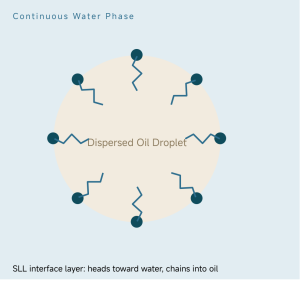
08 · Interface Engineering Across Formulation Structures
Oil/water interfaces: stabilization of emulsions and dispersions. Surfactant assemblies: complex cleansing structures, foam, and after-feel. Lipid-rich systems: dispersion and organization of ceramides, cholesterol, and other lipids.
SLL participates at multiple structural levels from adsorption to system organization.
09 · Mild Cleansing System Design
SLL’s anionic surfactant structure supports soil/sebum removal in combined systems, balancing cleansing with after-feel.
Formats: facial cleansers, body/hand washes, bathing products, and mild-cleansing concepts.
Patent context: cleansing, moisturization, lather, and skin feel. WO1998027193A1, Personal cleansing bar composition containing sodium lauroyl lactylate (1998); WO2007100784A2, Acyl lactylate compositions for rinse-out and leave-on applications for skin and hair (2007).
10 · From Clean to Comfortable Clean
The source compares conventional clean/tight/dry after-feel with an SLL formulation concept of clean/soft/comfortable/refined after-feel.
Published formulation goals: skin softness, moisturization, creamy lather, and improved after-feel. These reflect combined formulations, not effects attributable entirely to one ingredient.
Patent sources: WO1998027193A1; WO2007100784A2.
11 · An Interface Partner for Barrier Lipid Formulations
Ceramides + Cholesterol + Fatty Acids + SLL interface support.
Roles: dispersion, interface activity, co-emulsification, and lipid-delivery design. The lipid matrix is schematic.
US20240000681A1 (2024) includes SLL in designs containing Ceramide NP/AP/EOP, cholesterol, and free fatty acids/precursors. This is a patent disclosure, not PuriActives® SLL clinical data.
Patent title: Chemical compositions providing long-lasting skin pH control for improving skin-barrier function and treatment of skin-barrier disorders.
12 · Barrier-Friendly Formulation Architecture
SLL supports the architecture of lipid-based barrier-care products.
Combination logic: SLL interface support + Ceramides + Cholesterol + Phytosphingosine + Fatty Acids + Humectants.
Formats: barrier cream, ceramide lotion, sensitive/dry skin care, post-cleansing care, and lipid-rich serum. This formulation rationale does not by itself demonstrate clinical barrier repair.
13 · Moisturization Starts at the Interface
Hydration involves humectancy, occlusion, lipid organization, cleansing mildness, and emulsion quality.
SLL primarily supports interface, lipid, and cleansing dimensions of comfortable moisturization systems.
Patent context: skin softness, moisturization, cleansing, and foaming. WO2007100784A2 (2007); WO1998027193A1 (1998).
14 · From Skin to Hair
One molecule supports skin/hair formulation contexts. Published directions include shampoo, mild shampoo, scalp cleansing, rinse-off care, leave-on hair/scalp concepts, and hair grooming products.
15 · From Foam Quantity to Foam Quality
EP0562720A1, Hair shampoo (1993), examines acyl lactylates with alkyl polyglucosides (APG), including Sodium Lauroyl Lactylate-related systems, to address coarse APG foam.
Patent example range: acyl lactylates 3–15%, preferred 4–8%. These are patent-system ranges, not PuriActives® SLL recommendations.
Fine-Tune the Foam. Elevate the Cleansing Experience.
16 · Published Shampoo Formulation Examples
WO2007100784A2 (2007): approximately 3.0% SLL in a clear mild shampoo example concerned with cleansing, foam, viscosity, and pH stability; approximately 2.0% in another shampoo example for mild cleansing/sensory performance.
These are published examples, not recommended use levels. SLL has a documented hair-cleansing formulation history. Relevant attributes: cleansing, foaming, viscosity, pH stability, and sensory properties.
17 · Hair Fiber and Scalp Interface
Scalp: cleansing-system design, sebum/soil removal, freshness, mild formulations, and lipid-compatible care.
Hair: cleansing sensory properties, softer after-feel concepts, creamy foam, lipid-containing formulations, and conditioner/mask support.
Cosmetic scope excludes hair growth, anti-hair-loss, follicle repair, and hair-thickening claims.
18 · SLL and Modern Hair-Care Concepts
Reduced-sulfate cleansing, APG shampoos, scalp barrier care, ceramide hair care, lipid-replenishing shampoos, sensitive-scalp cleansing, premium cream shampoos, and scalp/hair serum emulsions.
An interface ingredient bridging cleansing technology and lipid care.
19 · SLL Interactions with Model Lipid Membranes
Gooran N, Tan SW, Yoon BK, Jackman JA. Int J Mol Sci. 2023;24(11):9283. DOI: 10.3390/ijms24119283.
Methods: QCM-D, quartz crystal microbalance with dissipation monitoring; EIS, electrochemical impedance spectroscopy; SLB, supported lipid bilayer; tBLM, tethered bilayer lipid membrane.
The study tracked SLL insertion, reorganization, and disruption of model membranes. The schematic shows aqueous phase, SLL, membrane perturbation, and solid support. Model lipid membranes are not equivalent to the human stratum corneum.
Source link: https://doi.org/10.3390/ijms24119283
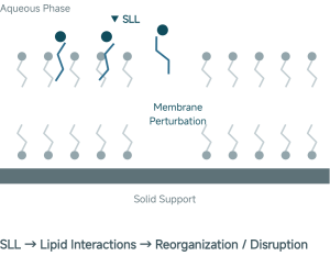
20 · Research Possibilities Beyond Emulsification
Lactylate membrane-interactive behavior offers research perspectives for cleansing and lipid-care design.
Directions: advanced cleansing, scalp hygiene, personal cleansing, microbial membrane research, and functional surfactant systems. These are research concepts without therapeutic claims.
Source: Gooran et al. 2023.
21 · Application Matrix
Skin: facial cleanser, cream cleanser, body wash, hand wash, moisturizing lotion, barrier cream, ceramide cream, facial emulsion, sensitive-skin care, lipid-rich serum, body lotion, and post-cleansing care.
Hair: shampoo, mild/cream shampoo, scalp cleanser/care, conditioner, mask, hair milk, leave-on emulsion, ceramide hair care, and lipid-replenishing care.
22 · Combination Design
Surfactants: APG, amphoteric, anionic, and nonionic systems. Lipids: ceramides, cholesterol, fatty acids, vegetable oils, and esters. Conditioning: glycerin, panthenol, hyaluronic acid, phytosphingosine, niacinamide, proteins, and amino acids.
Formulation versatility requires experimental compatibility testing; not every combination is presumed compatible.
23 · pH and Formulation Window
Published patent window: pH 4.0–8.5; preferred pH 5.0–7.0. The scale shown spans 3–9.
These are WO2007100784A2 acyl-lactylate skin/hair formulation windows, not product specifications.
pH 5–7 informs mildly acidic skin-care, gentle scalp-care, and hair-cleansing concepts.

24 · Reported Cosmetic Use
2024 CIR safety-assessment reported maximum use concentrations: 10% in rinse-off skin cleansing; 6.1% in leave-on body/hand products. These are reported concentrations, not product recommendations.
Chinese historical-use references in the PPT: approximately 10% rinse-off and 1.25% leave-on. Historical use is not a recommended level; the latest official inventory/public information governs interpretation.
The source describes peer-reviewed assessment, multiple categories/concentrations, Chinese use records, and rinse-off/leave-on applications.
Sources: Johnson et al. Int J Toxicol. 2024; Chinese ingredient-inventory public-use information. Regulatory/database statements are retained from the supplied PPT and have not been independently updated for this conversion.
25 · Safety Assessment Foundation
Johnson W Jr, Bergfeld WF, Belsito DV, et al. Safety Assessment of Alkanoyl Lactyl Lactate Salts as Used in Cosmetics. Int J Toxicol. 2024;43(4 Suppl). DOI: 10.1177/10915818241259694.
The source summarizes the Expert Panel conclusion as safe under current uses/concentrations when formulated to be nonirritating and nonsensitizing. This assessment includes the family containing Sodium Lauroyl Lactylate.
Foundation: established use, peer-reviewed assessment, rinse-off/leave-on contexts, and INCI recognition. This is a formulation-safety framework.
Source link: https://doi.org/10.1177/10915818241259694
26 · One Ingredient, Multiple Formulation Advantages
Emulsify: oil/water interface management. Cleanse: mild-system design. Stabilize: uniform dispersions. Condition: skin/hair sensory properties. Lipid compatibility: support for barrier-lipid formulations. Skin + Hair: dual application.
PuriActives® SLL integrates emulsifier, surfactant, and lipid-system support functions in a lactylated amphiphilic molecule.
27 · More Than an Emulsifier
Smarter interface management. Better cleansing/care sensory experience. Broader possibilities from skin to hair.
PuriActives® SLL: The Lactylate Interface for Modern Skin & Hair Care.
PuriPharm Co., Ltd. INCI: SODIUM LAUROYL LACTYLATE.
28 · Scientific References
Peer-reviewed literature, patent publications, databases, and regulatory sources from the presentation are retained below. Patents list publication numbers, titles, and years; applicant information is outside the source material.
Source link: https://doi.org/10.3390/ijms24119283
Source link: https://doi.org/10.1177/10915818241259694
Source link: https://pubchem.ncbi.nlm.nih.gov/compound/Sodium-lauroyl-lactylate
Source link: https://ec.europa.eu/growth/tools-databases/cosing/
PEER-REVIEWED LITERATURE
1. Gooran N, Tan SW, Yoon BK, Jackman JA. Unraveling Membrane-Disruptive Properties of Sodium Lauroyl Lactylate and Its Hydrolytic Products: A QCM-D and EIS Study. International Journal of Molecular Sciences. 2023;24(11):9283. DOI: 10.3390/ijms24119283
2. Johnson W Jr, Bergfeld WF, Belsito DV, et al. Safety Assessment of Alkanoyl Lactyl Lactate Salts as Used in Cosmetics. International Journal of Toxicology. 2024;43(4 Suppl). DOI: 10.1177/10915818241259694
PATENTS
WO2007100784A2 (2007)— Acyl lactylate compositions for rinse-out and leave-on applications for skin and hair.
EP0562720A1 (1993)— Hair shampoo.
WO1998027193A1 (1998)— Personal cleansing bar composition containing sodium lauroyl lactylate.
US20240000681A1 (2024)— Chemical compositions providing long-lasting skin pH control for improving skin-barrier function and treatment of skin-barrier disorders.
DATABASES & REGULATORY SOURCES
PubChem — Sodium Lauroyl Lactylate
EU CosIng — Sodium Lauroyl Lactylate
Cosmetic Ingredient Review / Expert Panel for Cosmetic Ingredient Safety
Current Chinese Inventory of Existing Cosmetic Ingredients and official public ingredient-use information
Patents list publication numbers, titles, and years; applicant information is outside this material.
NanoActive™ 10-HDA
01 · NanoActive™ 10-HDA: Nano-Encapsulated Royal Jelly Acid
Advanced liposomal active ingredient. Powered by PURISOME® FlexVes™ flexible-vesicle liposome technology. Royal Jelly, Re-Engineered. PuriPharm Co., Ltd.
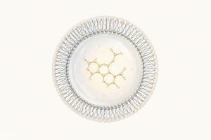
02 · The Royal Jelly Signature Molecule
Royal jelly contains proteins, sugars, lipids, amino acids, and trace components. 10-HDA is a characteristic fatty acid and a marker in research and quality evaluation. It provides a defined molecular active beyond the traditional natural-ingredient concept.
Identity: 10-Hydroxy-2-Decenoic Acid; 10-HDA/10-H2DA; Queen Bee Acid; Royal Jelly Acid; a medium-chain ω-hydroxy unsaturated fatty acid and characteristic royal jelly lipid.
10-HDA is a multifunctional bioactive lipid. Whole-royal-jelly effects cannot all be attributed to it; this presentation focuses on the lipid’s evidence.
03 · From Royal Jelly to 10-HDA
Traditional natural origin, molecular precision, and nano delivery.
1 Royal jelly, a traditional natural ingredient. 2 Identify the signature bioactive lipid. 3 Define molecular 10-HDA. 4 Study multiple skin biological pathways. 5 NanoActive™ liposomal delivery.
Applications: premium skin and scalp care. Defined. Studied. Delivered.

04 · 10-HDA Molecular Science
2D structure source: PubChem CID 5312738.
Formula C10H18O3; molecular weight approximately 186.25. Structure: α,β-unsaturated carboxylic acid, terminal hydroxyl, and a medium-chain fatty acid backbone. Class: medium-chain ω-hydroxy unsaturated fatty acid.
Potential: lipid interactions with skin lipids; hydroxyl-mediated hydrogen bonding; amphiphilic biofilm interactions; and a structural basis for epidermal biology research.
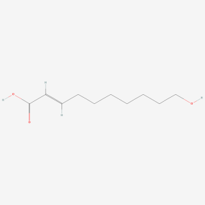
05 · One Royal Jelly Lipid, Multiple Skin Pathways
Seven research directions: barrier support, moisture/NMF, environmental stress defense, collagen/photoaging, skin tone, microbiome support, and soothing.
The presentation organizes an evidence network around one defined bioactive lipid. This mechanism map does not establish finished-product clinical efficacy.
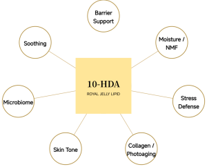
06 · Filaggrin: From 10-HDA to Natural Moisturizing Factors
Gu, Zeng, and Maeda (2017) found induction of filaggrin and free amino acids in cultured 3D human epidermis. This extends hydration concepts into cornification and NMF biology.
Key findings: increased filaggrin expression, FLG mRNA/protein, stratum-corneum free amino acids, and support for NMF formation.
Pathway: 10-HDA, increased filaggrin, free amino acids, NMF reserves, and moisture-retention/barrier appearance concepts.
Source: Cosmetics. 2017;4(4):48. DOI: 10.3390/cosmetics4040048. In vitro 3D epidermis.
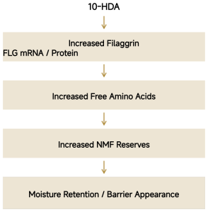
07 · Barrier Mechanism Map
From keratinocytes to the stratum corneum: differentiation support, increased granular-layer filaggrin, amino acid release, NMF reserves, hydration, and a stronger-looking barrier.
Layer schematic: stratum corneum, NMF/hydration; stratum granulosum, filaggrin processing and amino acids/NMF; keratinocytes, differentiation/cornification and the proposed 10-HDA starting point.
Source: Gu et al. Cosmetics. 2017;4(4):48. DOI: 10.3390/cosmetics4040048. The diagram illustrates mechanisms and does not establish product penetration depth.
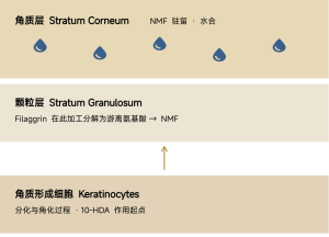
08 · NQO1: Supporting Epidermal Stress Defense
Okumura et al. (2021) identified 10-HDA as an important royal jelly fatty acid inducing NQO1, an antioxidant/detoxification enzyme.
Care contexts: UV oxidative stress, ROS accumulation, pollution/environmental stress, and early oxidative-aging signs. The positioning is defense support, without sunscreen or medicinal claims.
Pathway: 10-HDA, cellular stress-response signals, increased NQO1, enhanced antioxidant defense, and reduced oxidative-stress burden.
Source: Int J Mol Sci. 2021;22(23):12973. DOI: 10.3390/ijms222312973. In vitro cellular/epidermal models.
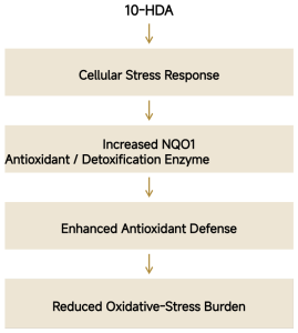
09 · 10-HDA and UVA-Induced Dermal Damage
Human dermal fibroblast pathway: UVA, increased ROS, JNK/p38 signaling, increased MMP-1/MMP-3, and collagen breakdown/photoaging.
10-HDA findings: lower ROS and senescence-related changes, modulated MMP-1/MMP-3, supported type I collagen expression, and protection against UVA cellular damage.
Zheng et al. (2013) connect ROS management with downstream MMP modulation and collagen protection.
Source: J Eur Acad Dermatol Venereol. 2013;27:1269–1277. DOI: 10.1111/j.1468-3083.2012.04707.x. In vitro human fibroblasts.
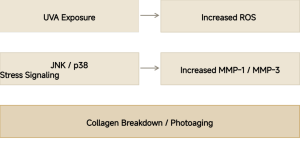
10 · Royal Jelly Biology and Collagen Science
This section concerns whole royal jelly, separate from purified 10-HDA.
Park et al. (2011), human skin fibroblast UVB model: royal jelly enhanced collagen production, with TGF-β1-related responses, type I procollagen, and extracellular-matrix support.
Concept: protect and preserve youthful-looking skin. Whole royal jelly and 10-HDA findings inform complementary collagen narratives, but whole-royal-jelly results cannot be fully attributed to 10-HDA. The source schematic includes Royal Jelly/10-HDA; the experiment itself tested whole royal jelly.
Source: J Med Food. 2011;14(9):899–906. DOI: 10.1089/jmf.2010.1363. In vitro.
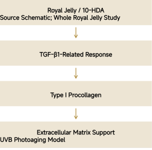
11 · 10-HDA and Melanogenesis
Peng et al. (2017) studied B16F1 melanoma cells and topical mouse skin, showing melanogenesis inhibition involving MITF, Tyrosinase, and TRP pathways.
Models assessed melanin/tyrosinase and topical tone-related indicators. Multi-target concepts support clearer-looking tone and appearance; no pigment-disease treatment claims.
Pathway: 10-HDA inhibition, MITF modulation, Tyrosinase, TRP-1/TRP-2, and melanin synthesis.
Source: BMC Complement Altern Med. 2017;17:392. DOI: 10.1186/s12906-017-1888-8.
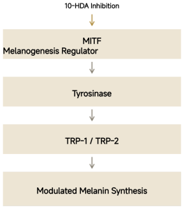
12 · Staphylococcus aureus Biofilm Research
In vitro findings: reduced biofilm formation, EPS production, adhesion, aggregation, and mature-biofilm viability.
Genes: reduced sarA, agrA, and hla, associated with biofilm regulation, quorum signaling, and α-hemolysin virulence.
Positioning: a microbiome-friendly skin-purifying strategy managing unfavorable biofilms and environments. Care concepts include oily/blemish-prone skin, sensitive-skin microbiome care, scalp care, skin cleansing care, and fragile barriers. These findings do not establish selective support for all beneficial species.
Gao et al. Molecules. 2022;27(5):1485. DOI: 10.3390/molecules27051485.
13 · The Delivery Challenge
01 Poor aqueous solubility limits direct water-based use. 02 Limited oil solubility constrains conventional oil delivery. 03 Free-state penetration depends on dosage-form design. 04 Direct addition can be difficult to disperse and restrict formulation options.
Biological activity does not by itself establish bioavailability at the intended interface.
14 · PURISOME® FlexVes™: Why Liposomes
A lipid-bilayer vesicle is a membrane-related carrier architecture for loading/dispersing 10-HDA. It aims to improve aqueous dispersion, compatibility with stratum-corneum lipids, and delivery/deposition potential.
Values: nanoscale delivery, lipid compatibility, dispersion, skin-deposition potential, controlled skin interactions, and formulation flexibility.
The vesicle image is a mechanism schematic, not measured particle-size or encapsulation-efficiency data.
15 · 2026 Independent Nano-Delivery Evidence
Gao et al. (2026) studied Hyalurosomes encapsulating a 10-HDA/matrine deep eutectic solvent, using ex vivo skin and cellular models.
Formulation/delivery: improved dissolution/dispersion and stability, with increased deposition in stratum corneum, viable epidermis, and dermis.
Cells: reduced HaCaT ROS, IL-1α/IL-6/TNF-α; increased HaCaT migration; reduced HFF-1 senescence phenotype.
The formulation is distinct from NanoActive™ 10-HDA. It supports the delivery strategy and does not constitute direct product evidence.
Int J Pharm. 2026;701:127150. DOI: 10.1016/j.ijpharm.2026.127150.
16 · Free and Nano-Encapsulated 10-HDA
Free 10-HDA: limited solubility, dispersion, penetration, and formulation choices.
NanoActive™ 10-HDA with PURISOME® FlexVes™: liposomal dispersion, a skin-compatible lipid carrier, improved delivery potential, greater formulation versatility, and modern skin-care design.
Better delivery offers greater opportunity for biological activity; product performance still requires direct testing.
17 · Integrated Mechanism Network
Epidermis: filaggrin, amino acids/NMF, moisture/barrier. Dermis: lower ROS/MMP-1/MMP-3, collagen preservation. Stress defense: NQO1 and oxidative-defense support. Pigmentation: MITF/Tyrosinase/TRP and even-looking tone. Microbiome: biofilm/virulence modulation and skin-environment concepts.
One bioactive lipid, multiple pathways. The network combines Gu 2017, Okumura 2021, Zheng 2013, Peng 2017, and Gao 2022, retaining their original model-level attribution.
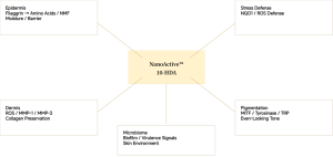
18 · Five Core Skincare Values
01 Barrier recovery: filaggrin/NMF and hydration foundations. 02 Anti-stress defense: NQO1-related epidermal defenses. 03 Photoaging care: ROS/MMP and collagen protection. 04 Tone: MITF/Tyrosinase-related pathways for dull/uneven appearance. 05 Microbiome: unfavorable-biofilm management and skin-environment support.
Barrier × Defense × Collagen × Tone × Microbiome connects mechanisms to formulation concepts.
19 · Scalp Is Skin
Scalp pressures: fragile barrier/moisture loss, oxidative stress, microbial imbalance, inflammatory environments, and sebum/pollution.
Skin mechanisms inform scalp concepts: Filaggrin/NMF for hydration; NQO1 for stress defense; biofilm management for microbial care; soothing pathways for comfort. These concepts require scalp-specific validation.
Healthy scalp, a better environment for beautiful hair. The positioning does not claim direct hair growth.
20 · Royal Jelly and Scalp Research Clues
A 2024 clinical observation of a medicated hair tonic containing royal jelly extract associated the combination product with improved scalp moisture/condition and hair condition. Dermal papilla experiments reported changes in some growth-related genes.
Other actives were present; results cannot all be attributed to royal jelly extract or 10-HDA.
NanoActive™ positioning: scalp barrier, moisture, soothing, antioxidant defense, and microbiome care. Skin health guides scalp ecological concepts.
J Soc Cosmet Chem Jpn. 2024;58(1):55–64. DOI: 10.5107/sccj.58.55. Combination-product clinical observation and cell experiments.
21 · Skincare Applications
Anti-aging serum, repair serum, barrier cream, sensitive-skin serum, overnight recovery, tone-care serum, blemish-prone care, microbiome products, urban-defense serum, eye-contour care, premium royal jelly concepts, and post-treatment-inspired care.
Royal Jelly Renewal Serum: photoaging/collagen concept. Barrier Defense Cream: Filaggrin–NMF hydration concept. Bio-Balance Repair Essence: biofilm-management concept.
22 · Scalp and Hair Applications
Scalp serum, ampoule, shampoo, soothing mist, dry-scalp lotion, oily-scalp essence, post-color/perming scalp care, anti-pollution serum, and premium aging scalp care.
Royal Scalp Defense Serum: antioxidant/environmental defense. Scalp Barrier Booster: daily barrier/hydration care. Nano Royal Jelly Scalp Essence: premium nano-delivery and microbiome concepts.
23 · Value for Formulators
01 Easier incorporation addresses free-active solubility, dispersion, and penetration challenges. 02 Lipid-compatible delivery. 03 Water-based formulation design for serums/lotions. 04 Multiple barrier, antioxidant, aging, tone, and microbiome concepts. 05 Premium storytelling: Royal Jelly × Molecular Science × Nano Delivery.
Nature × Molecule × Nanotechnology combines natural-origin narrative, molecular evidence, and delivery design.
24 · Royal Jelly, Re-Engineered
01 A signature royal jelly lipid. 02 Filaggrin/NMF hydration. 03 Epidermal defense and collagen-protection research. 04 Microbiome/tone concepts. 05 Advanced liposomal design to address practical delivery barriers.
NanoActive™ 10-HDA, powered by PURISOME® FlexVes™.
25 · Positioning Matrix
Traditional royal jelly: a strong natural narrative with complex composition. Free 10-HDA: a defined molecule with formulation/delivery challenges. NanoActive™ 10-HDA: Defined Bioactive + Nano Delivery; Molecular Precision + Advanced Delivery.
Horizontal axis: evolution of active form. Vertical axis: narrative/scientific level. The schematic positions the product at molecular precision and advanced delivery; it is a qualitative positioning map, not quantitative comparative data.
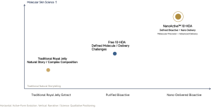
26 · References
All ten references from the presentation are retained below.
Molecular-structure source: PubChem CID 5312738. The science concerns 10-HDA (10-Hydroxy-2-Decenoic Acid, C10H18O3), distinct from 10-Hydroxydecanoic Acid (10-HDAA). Final INCI follows the formal PuriPharm Co., Ltd. TDS/SPEC.
PuriPharm Co., Ltd. Royal Jelly, Re-Engineered.
1. Gu L, Zeng H, Maeda K. 10-Hydroxy-2-Decenoic Acid in Royal Jelly Extract Induced Both Filaggrin and Amino Acid in a Cultured Human Three-Dimensional Epidermis Model. Cosmetics. 2017;4(4):48. DOI: 10.3390/cosmetics4040048.
2. Zheng J, Lai W, Zhu G, et al. 10-Hydroxy-2-Decenoic Acid Prevents Ultraviolet A-Induced Damage and Matrix Metalloproteinases Expression in Human Dermal Fibroblasts. J Eur Acad Dermatol Venereol. 2013;27:1269–1277. DOI: 10.1111/j.1468-3083.2012.04707.x.
3. Park HM, Hwang E, Lee KG, et al. Royal Jelly Protects Against Ultraviolet B-Induced Photoaging in Human Skin Fibroblasts via Enhancing Collagen Production. J Med Food. 2011;14(9):899–906. DOI: 10.1089/jmf.2010.1363.
4. Peng CC, Sun HT, Lin IP, et al. The Functional Property of Royal Jelly 10-Hydroxy-2-Decenoic Acid as a Melanogenesis Inhibitor. BMC Complement Altern Med. 2017;17:392. DOI: 10.1186/s12906-017-1888-8.
5. Okumura N, Ito T, Degawa T, et al. Royal Jelly Protects against Epidermal Stress through Upregulation of the NQO1 Expression. Int J Mol Sci. 2021;22(23):12973. DOI: 10.3390/ijms222312973.
6. Gao K, Su B, Dai J, et al. Anti-Biofilm and Anti-Hemolysis Activities of 10-Hydroxy-2-Decenoic Acid against Staphylococcus aureus. Molecules. 2022;27(5):1485. DOI: 10.3390/molecules27051485.
7. Perminaite K, Fadda AM, Sinico C, Ramanauskiene K. Formulation of Liposomes Containing Royal Jelly and Their Quality Assessment. DOI: 10.1166/jnn.2021.19053.
8. Gao C, Liu F, Yang S, Wang Q, Yang C, Liang R. 10-Hydroxy-2-Decenoic Acid/Matrine Deep Eutectic Solvent Encapsulated in Hyalurosomes for Enhanced Transdermal Delivery and Antioxidant Efficacy. Int J Pharm. 2026;701:127150. DOI: 10.1016/j.ijpharm.2026.127150.
9. Enhanced Skin Penetration of 10-Hydroxy-2-Decenoic Acid via Anionic Polymer Formulations and Skin Hydration Modulation. J Dermatologic Sci Cosmet Technol. 2026;3(1):100144. DOI: 10.1016/j.jdsct.2025.100144.
10. The Effectiveness of Medicated Hair Tonic Containing Royal Jelly Extract. J Soc Cosmet Chem Jpn. 2024;58(1):55–64. DOI: 10.5107/sccj.58.55.